FIGURE SUMMARY
Title

SLC41A1 is essential for magnesium homeostasis in vivo

Authors
Arjona, F.J., Latta, F., Mohammed, S.G., Thomassen, M., van Wijk, E., Bindels, R.J.M., Hoenderop, J.G.J., de Baaij, J.H.F.
Source
Full text @ Pflügers Archiv. / Eur. J. Physiol.

ZFIN is incorporating published figure images and captions as part of an ongoing project. Figures from some publications have not yet been curated, or are not available for display because of copyright restrictions.

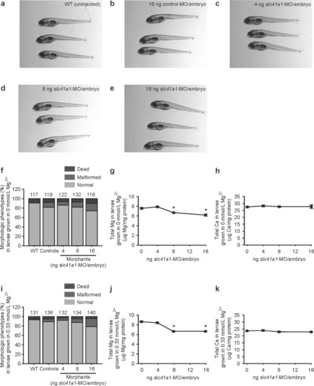
Slc41a1 knockdown evokes renal Mg2+ wasting in zebrafish. ae Representative images of the normal morphologic phenotypes obtained in untreated (WT) 120 hpf zebrafish grown in Mg2+-free E3 medium and after injection of 16 ng control-MO/embryo or 4–16 ng slc41a1-MO/embryo (splice-site blocking MO). f Distribution of morphologic phenotypes in zebrafish larvae (120 hpf) untreated (WT) or after injection with different doses of slc41a1-MO (splice-site blocking MO) or control-MO. Fish were grown in Mg2+-free E3 medium. Numbers on top of the bars indicate the number of animals in each experimental condition. g, h Total Mg (g) and Ca (h) content in morphologically normal 120 hpf zebrafish grown in Mg2+-free E3 medium after injection with different doses of slc41a1-MO (splice-site blocking MO); the dose of zero represents injection with 16 ng control-MO/embryo (n = 10). i Distribution of morphologic phenotypes in zebrafish larvae (120 hpf) untreated (WT) or after injection with different doses of slc41a1-MO (splice-site blocking MO) or control-MO. Fish were grown in E3 medium (0.33 mmol/L Mg2+). Numbers on top of the bars indicate the number of animals in each experimental condition. j, k Total Mg (j) and Ca (k) content in morphologically normal 120 hpf zebrafish grown in E3 medium (0.33 mmol/L Mg2+) after injection with different doses of slc41a1-MO (splice-site blocking MO); the dose of zero represents injection with 16 ng control-MO/embryo (n = 10). g, h, j, k Data are presented as mean ± S.E.M. Asterisks indicate significant differences between control and experimental groups (*P < 0.05)

ZFIN is incorporating published figure images and captions as part of an ongoing project. Figures from some publications have not yet been curated, or are not available for display because of copyright restrictions.

ZFIN is incorporating published figure images and captions as part of an ongoing project. Figures from some publications have not yet been curated, or are not available for display because of copyright restrictions.

EXPRESSION / LABELING:
Genes:
Fish:
Knockdown Reagent:
Anatomical Term:
Stage: Day 5
PHENOTYPE:
Fish:
Knockdown Reagent:
Observed In:
Stage: Day 5

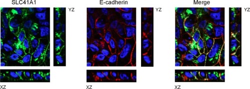
SLC41A1 is expressed at the basolateral membrane. Localization of SLC41A1 (green) and E-cadherin (red) in stably transfected polarized MDCKI cells with HA-tagged SLC41A1 is shown by immunofluorescence and confocal analyses in Z, XZ, and YZ sections. In blue, DAPI staining

Acknowledgments
This image is the copyrighted work of the attributed author or publisher, and ZFIN has permission only to display this image to its users. Additional permissions should be obtained from the applicable author or publisher of the image. Full text @ Pflügers Archiv. / Eur. J. Physiol.