
- Title
-
Cog4 is required for protrusion and extension of the epithelium in the developing semicircular canals
- Authors
- Clément, A., Blanco-Sánchez, B., Peirce, J.L., Westerfield, M.
- Source
- Full text @ Mech. Dev.
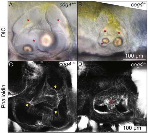
The pillars do not form properly in cog4−/− mutants. Live images of the inner ear of cog4+/+ sibling (A, n = 34 larvae) and cog4−/− mutant larvae (B, n = 15 larvae). Red stars indicate the semicircular canals. Phalloidin staining of the inner ear of cog4+/+ sibling (C, n = 24 larvae) and cog4−/− mutant larvae (D, n = 25 larvae). Yellow arrowheads indicate the fusion plates that form in 100% (24 out of 24) of wild-type siblings (C). Red arrowhead points to the malformed pillar. One or more malformed pillars are observed in 84% (21 out of 25) of homozygous mutant larvae (D). Anterior to the left and dorsal to the top. 5 dpf. PHENOTYPE:
|
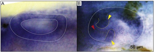
cog4 is differentially expressed during inner ear morphogenesis. In situ hybridization of cog4 in the ear at 28 hpf (A, n = 18 embryos), and 52 hpf (B, n = 18 larvae). Dorsal view, anterior to the left (A). Lateral view, anterior to the left (B). Yellow arrowheads indicate the anterior and ventral projections. Red arrowhead indicates the hair cells of the anterior macula (B). The inner ear is outlined in white (A, B) and the epithelial projections in yellow (B). |
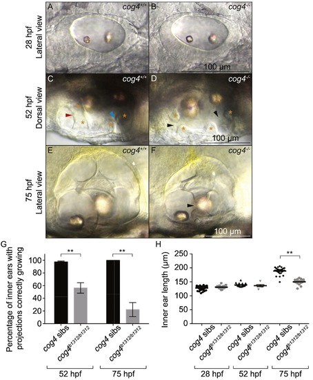
Formation of the epithelial projections is delayed in cog4−/−mutants. Live images of the inner ear from cog4+/+ sibling (A, n = 49 embryos; C, n = 57 larvae; E, n = 26 larvae) and cog4−/− mutant larvae (B, n = 21 embryos; D, n = 22 larvae; F, n = 18 larvae) at 28 hpf (A, B), 52 hpf (C, D) and 75 hpf (E, F). Orange stars indicate the anterior and posterior projections and bulges (C, D). The red and blue arrowheads indicate the anterior and forming posterior pillar, respectively (C). Black arrowheads point to the gaps between the anterior and forming posterior projections and bulges (D, F). Anterior to the left (A–F). Dorsal to the top (A, B, E, F). Medial to the top (C, D). Percentage of inner ears with projections growing correctly at 52 hpf and 75 hpf, in cog4+/+ sibling (52 hpf, n = 114 inner ears; 75 hpf, n = 26 inner ears) and cog4−/− mutant larvae (52 hpf, n = 38 inner ears; 75 hpf, n = 18 inner ears) (G). Ear size at 28, 52 and 75 hpf in cog4+/+ sibling (28 hpf, n = 49 embryos; 52 hpf, n = 18 larvae; 75 hpf, n = 26 larvae) and cog4−/− mutant larvae (28 hpf, n = 21 embryos; 52 hpf, n = 6 larvae; 75 hpf, n = 18 larvae) (H). Size was measured along the antero-posterior axis of the inner ear. Data are represented as mean ± SEM. **p < 0.01. PHENOTYPE:
|
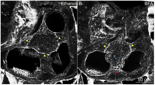
The pillars do not form properly in larvae treated with BFA. Phalloidin staining of the inner ear in larvae treated with ethanol (A; control, n = 10 larvae) or BFA (B, n = 10 larvae). Yellow arrowheadsindicate the fusion plates. Red arrowhead indicates an abnormal fusion plate. Blue star indicates the malformed pillar. Anterior to the left and dorsal to the top. 5 dpf. PHENOTYPE:
|
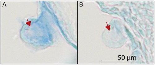
The secretion of proteoglycans from the epithelial projections is reduced in cog4−/− mutants. Paraffin section of the inner ear posterior projection in cog4+/+ (A, n = 6 larvae) and cog4−/− larvae (B, n = 7 larvae). Alcian blue staining was performed prior to sectioning. Red arrows indicate the projection core. Anterior to the left and dorsal to the top. 52 hpf. PHENOTYPE:
|
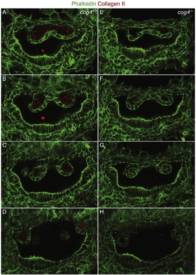
The secretion of Collagen type II from the epithelial projections is reduced in cog4−/− mutants. Series of confocal sections through the inner ear of cog4+/+ (A–D; n = 9 larvae) and cog4−/− larvae (E–H, n = 8 larvae). The most lateral section is at the top and most medial at the bottom. Anterior to the left and dorsal to the top. 52 hpf. PHENOTYPE:
|
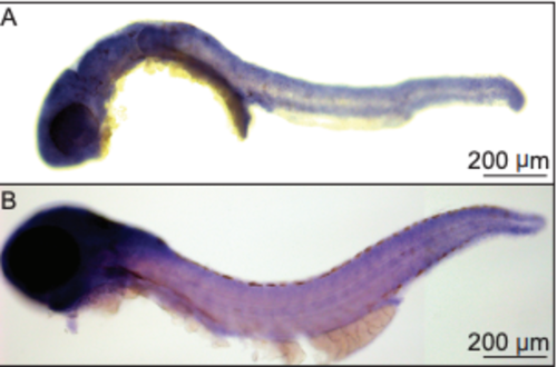
cog4 is ubiquitously expressed. In situhybridization of cog4 in WT whole embryo at 28 hpf (n = 18 embryos) and WT whole larvae at 52 hpf (n = 18 larvae). Anterior to the left and dorsal to the top. |
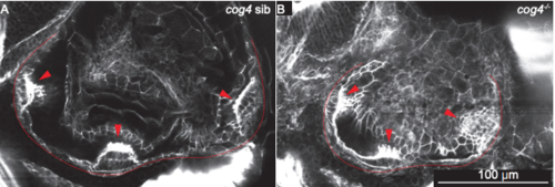
The three cristae form in cog4−/− mutants. Phalloidin staining of cog4 sibling (A, n = 23 larvae) and cog4−/−mutant larvae (B, n = 22 larvae). Red arrow heads point to the cristae and inner ear is outlined in red (A, B). Because of the inner ear shape in cog4−/− mutant larvae, the three cristae could not be imaged on the same exact plan. Anterior to the left and dorsal to the top. 5 dpf. PHENOTYPE:
|
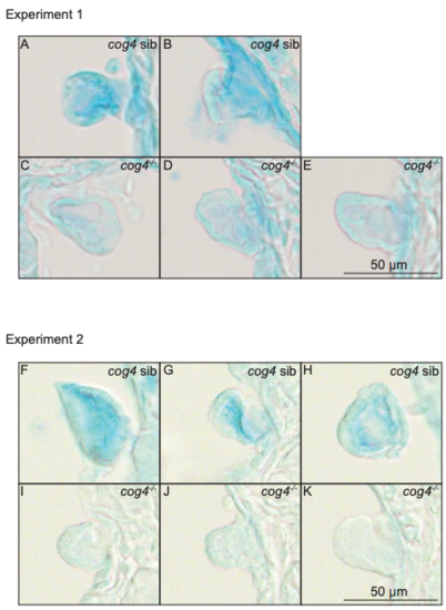
The secretion of proteoglycans from the epithelial projections is reduced in cog4−/− mutants. Paraffin sections of the inner ear anterior (C) and posterior projections (A, B, D, E, F–K) after Alcian blue staining in cog4+/+ (A, B, F–H; n = 6 larvae) and cog4−/− larvae (C–E, I–K; n = 7 larvae). Anterior to the left and dorsal to the top. 52 hpf. PHENOTYPE:
|
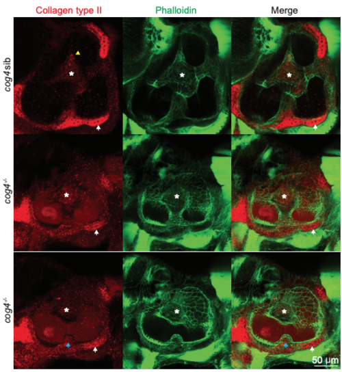
Collagen type II is present in the semicircular canal tissues of cog4−/− mutants. Co-labeling of Collagen type II (red) and phalloidin (green) in cog4 sibling (A; n = 22 larvae) and cog4−/− mutant larvae (B, C; n = 23 larvae). White stars indicate the pillars (A–C). White arrowheads point to the otic capsule (A–C). Blue arrowheads points to a delayed ventral projection (C). Anterior to the left and dorsal to the top. 5 dpf. PHENOTYPE:
|
Reprinted from Mechanisms of Development, 155, Clément, A., Blanco-Sánchez, B., Peirce, J.L., Westerfield, M., Cog4 is required for protrusion and extension of the epithelium in the developing semicircular canals, 1-7, Copyright (2018) with permission from Elsevier. Full text @ Mech. Dev.