
- Title
-
In vivo selectivity and localization of reactive oxygen species (ROS) induction by osmium anticancer complexes that circumvent platinum-resistance
- Authors
- Coverdale, J.P.C., Bridgewater, H.E., Song, J.I., Smith, N.A., Barry, N.P.E., Bagley, I., Sadler, P.J., Romero Canelon, I.
- Source
- Full text @ J. Med. Chem.
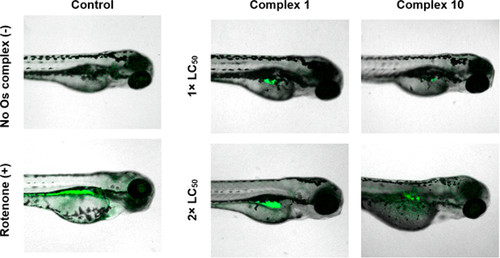
Reactive oxygen species (ROS) in anaesthetized whole-mount Singapore wild-type zebrafish (Danio rerio) treated with equipotent concentrations (1 or 2× LC50) of Os-sulfonamide 1 or Os-azopyridine 10 for 96 h. Fluorescence for ROS (green) is shown superimposed onto bright field images. Embryos were stained using the green reagent of the ROS/Superoxide Detection Kit (Enzo life sciences) to detect ROS. Excitation, 458 and 488 nm; green emission for ROS, 493–550 nm. Rotenone was the positive control; 50 μM, 2 min exposure.(47) |
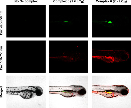
Two-color fluorescent imaging of whole mount SG-WT zebrafish (Danio rerio) treated with Os(II) sulfonamide 6 for 96 h. Fluorescence for ROS (green) and 6 (red) is shown superimposed onto bright field images. Overlapping regions (yellow) are shown. Confocal images were acquired using a Zeiss LSM880 confocal microscope. Embryos were stained using the green reagent of the ROS/Superoxide Detection Kit (Enzo life sciences) for ROS detection. Excitation, 458, 488, and 561 nm; green emission for ROS, 493–550 nm; red emission for 6, 568–750 nm. See SI for full confocal data. |
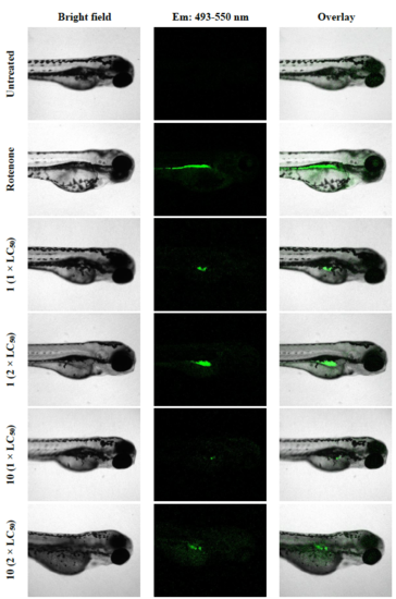
Reactive oxygen species (ROS) in anaesthetized whole-mount SGWT zebrafish (Danio rerio) treated with equipotent concentrations (1.0 or 2.0 × LC50) of Os-sulfonamide 1 or Os-azopyridine 10 for 96 h. Fluorescence for ROS (green) is shown superimposed onto bright field images. Confocal images were acquired using a Zeiss LSM880 confocal microscope. Embryos were stained using ROS Detection Kit (Enzo life sciences). Excitation: 458, 488 nm; green emission for ROS: 493-550 nm. Rotenone was the positive control (50 μM, 2 min). |