- Title
-
Reversibility of Proliferative Thyroid Lesions Induced by Iodine Deficiency in a Laboratory Zebrafish Colony
- Authors
- Murray, K.N., Wolf, J.C., Spagnoli, S.T., Lains, D., Budrow, N., Kent, M.L.
- Source
- Full text @ Zebrafish
|
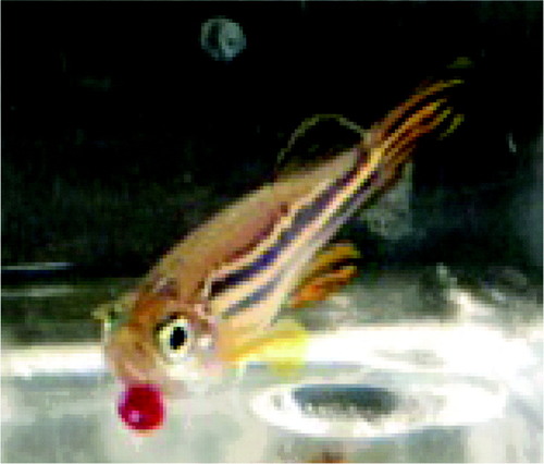
An adult zebrafish with a large red mass between the mandibles. |
|
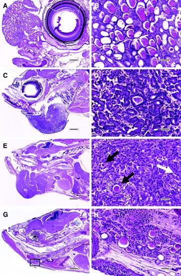
Thyroid lesions. (A) and (B) are ectopic follicular cell hyperplasia. (A) Hyperplastic lesion in the nasal region anterior to the eye. Follicular hyperplasia also occurred in the pharyngeal region and kidney of this fish to a lesser extent. H&E; scale bar, 250 μm. (B) Higher magnification of the lesion in A. H&E; scale bar, 50 μm. (C) and (D) are follicular cell adenoma. (C) The adenoma protrudes from the ventral jaw region, which was a common presentation for thyroid lesions in this report. H&E; scale bar, 500 μm. (D) Higher magnification of the lesion in (C). H&E; scale bar, 25 μm. (E) and (F) are follicular cell carcinoma. (E) This lesion also protrudes from the lower jaw, but compared to the adenoma in (C) and (D), this carcinoma is somewhat more irregular in outline and larger. H&E; scale bar, 800 μm. (F) Higher magnification of the lesion in (E). This tumor was characterized by sheets of cells with minimal follicle development (black arrow), cytologic atypia, and the presence of mitotic figures (white arrow). H&E; scale bar, 25 μm. (G) and (H) are recovering thyroid tissue. (G) Mass lesions are no longer present 5 months after iodine replenishment. The box indicates the location of (H). H&E; scale bar, 500 μm. (H) The thyroid tissue consists of numerous tiny follicles in the ventral jaw area. H&E; scale bar, 25 μm. H&E, hematoxylin and eosin. |


