FIGURE SUMMARY
- Title
-
Analysis of Beta-cell Function Using Single-cell Resolution Calcium Imaging in Zebrafish Islets
- Authors
- Janjuha, S., Pal Singh, S., Ninov, N.
- Source
- Full text @ J. Vis. Exp.
|
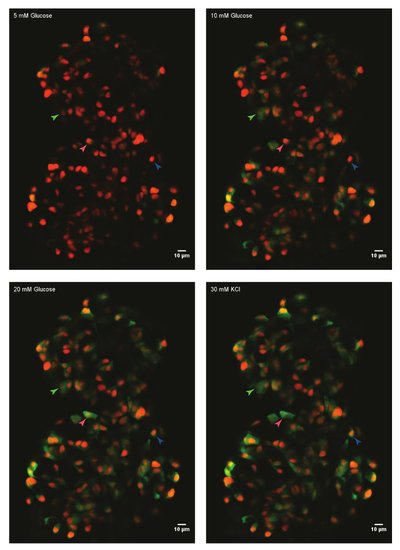
Ex vivo live-imaging of calcium influx using GCaMP6s in zebrafish beta-cells. A primary islet from Tg(ins:nls-Renilla-mKO2); Tg(ins:GCaMP6s) zebrafish (45 dpf) was mounted in fibrinogen-thrombin mold and incubated with 5 mM (basal) glucose. Beta-cells were labeled with a red nuclear marker, while the GCaMP6s fluorescence is present in the green channel. The islet was stimulated with a glucose-ramp consisting of sequential incubation with 10 mM and 20 mM D-glucose, and depolarized via the addition of 30 mM KCl. Arrowheads mark individual beta-cells whose activity was analyzed. |
Acknowledgments
This image is the copyrighted work of the attributed author or publisher, and
ZFIN has permission only to display this image to its users.
Additional permissions should be obtained from the applicable author or publisher of the image.
Full text @ J. Vis. Exp.

