
- Title
-
CRMP2 and CRMP4 Are Differentially Required for Axon Guidance and Growth in Zebrafish Retinal Neurons
- Authors
- Liu, Z.Z., Zhu, J., Wang, C.L., Wang, X., Han, Y.Y., Liu, L.Y., Xu, H.A.
- Source
- Full text @ Neural Plast.
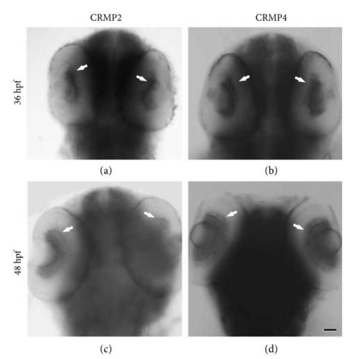
CRMP2 and CRMP4 are expressed in the retinal ganglion cell layer when retinal axons are exiting the eye and crossing the midline. Whole mount in situ hybridization was performed in zebrafish embryos using RNA probes for CRMP2. (a, b) CRMP2 and CRMP4 transcripts are detected in the retina (white arrows) at 36 hpf. (c, d) CRMP2 and CRMP4 transcripts are detected in the retina of 48 hpf embryos (white arrows). Scale bar: 50 μm. |
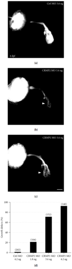
Knocking down CRMP2 induces growth defects of retinal axons. Morpholino was injected into the zygotes at the 1-2 cell stage. Embryos were allowed to grow until 4 days (4 days postfertilization, 4 dpf) and fixed with PFA. Lipophilic fluorescent dye DiI or DiD was injected into an eye of the larvae to label retinal axons. (a) An example showing that, in control MO-treated zebrafish larvae, retinal axons exit the eye, cross the midline, and grow into and arborize the whole tectum at 4 dpf. (b, c) Representative images of retinal axons of CRMP2 MO-treated embryos. Much less retinal axons grow into and arborized the tectum (white arrowheads) compared with that in control MO-treated embryos. (d) The growth defects of retinal axons induced by CRMP2 MOs are dose-dependent. The y-axis represents the percentage of eyes with growth defects of retinal axons. The doses of MOs are labeled under each column. The numbers in parentheses above each column indicate the amount of eyes. Scale bar: 50 μm. PHENOTYPE:
|
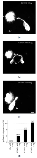
Knocking down CRMP4 causes ipsilateral misprojections of retinal axons. Morpholino was injected into 1-2 cell stage embryos and retinal axons were labeled with DiI or DiD at 4 dpf. (a) A representative image demonstrating that, in control MO-treated zebrafish larvae, all retinal axons cross the midline and project into the opposite side of the tectum. (b, c) Representative images of retinal axon guidance errors in CRMP4 MO-treated larvae. A part or all of the retinal axons fail to cross the midline and misproject into the ipsilateral tectum (arrows). Note that although the axons misproject ipsilaterally, they still follow the normal optic tract and arborize into the tectum. (d) The ipsilateral misprojections of retinal axons caused by CRMP4 MOs are dose dependent. The doses of MOs are labeled under each column. The numbers in parentheses above each column indicate the amount of eyes. Scale bar: 50 μm. PHENOTYPE:
|
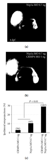
CRMP4 and Nrp1a synergize in retinal axon guidance. A low dose of either CRMP4 or Nrp1a induce a small percentage of ipsilateral misprojections. A half dose of either CRMP4 MOs or Nrp1a MOs was injected singly or in combination. (a, b) Representative images of knocking down effects by a half dose of Nrp1a MOs or Nrp1a and CRMP4 in combination. Some retinal axons fail to cross the midline and misproject ipsilaterally (arrows). (c) The combination of the two morpholinos induce a significantly higher percentage of ipsilateral misprojections than simply adding up the misprojections caused by the two half doses (Fisher’s exact test, ). The doses of MOs are labeled under each column. The numbers in each column indicate the amount of eyes. PHENOTYPE:
|
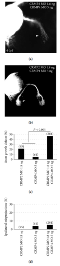
CRMP2 synergized with CRMP4 in axon growth but not axon guidance. A half dose of either CRMP2 or CRMP4 MOs was injected singly or in combination. (a, b) Representative examples of knocking down effects of CRMP2 and CRMP4 MOs in combination. (c) The combination of the two MOs causes a significantly higher percentage of axon growth defects than the sum of the defects caused by adding up the single half doses (Fisher’s exact test, ). (d) No obvious synergy between the two CRMPs in retinal axon guidance. The doses of MOs are labeled under each column. The numbers in parentheses above each column indicate the amount of eyes. PHENOTYPE:
|