
- Title
-
Small molecule inhibition of RAS/MAPK signaling ameliorates developmental pathologies of Kabuki Syndrome
- Authors
- Tsai, I.C., McKnight, K., McKinstry, S.U., Maynard, A.T., Tan, P.L., Golzio, C., White, C.T., Price, D.J., Davis, E.E., Amrine-Madsen, H., Katsanis, N.
- Source
- Full text @ Sci. Rep.

ZFIN is incorporating published figure images and captions as part of an ongoing project. Figures from some publications have not yet been curated, or are not available for display because of copyright restrictions. PHENOTYPE:
|
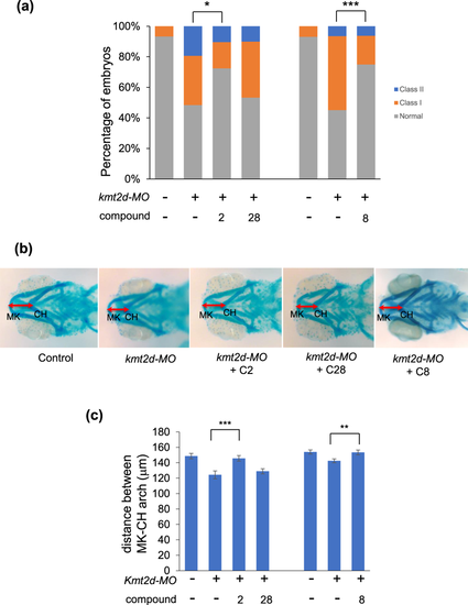
Compounds 2 and 8 ameliorate CE and jaw defects caused by loss of kmt2d. (a) Compounds 2 and 8, but not 28 (negative control), rescue CE defects of kmt2d morphants. (b) Alcian blue staining of jaw. Depletion of kmt2d leads to a change in jaw layout, reducing the distance between Meckel’s (MK) cartilage and ceratohyal (CH) cartilage (red double arrow). Treatment with compound 2 or 8, but not 28, ameliorates this defect in kmt2d morphants. (c) Quantitative measurement of the distance between MK and CH cartilages (n > 25). *p < 0.05; **p < 0.01; ***p < 0.001. Error bars show SEM (standard error of the mean). |
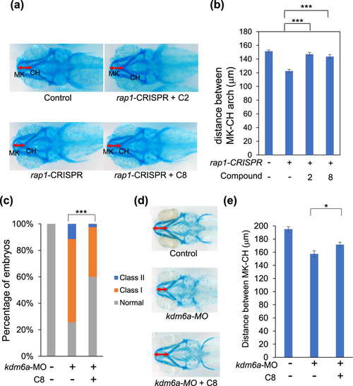
Compound 8 ameliorates the CE and jaw defects caused by loss of rap1 and kdm6a. (a) gRNA targeting both zebrafish rap1a and rap1b were injected into embryos at the 1-cell stage with Cas9 protein. Larvae were incubated with compound 8 (100 nM) at the 8-cell stage and scored for micrognathia. 5 dpf larvae were stained with Alcian blue to assess the layout of the jaw cartilage. In comparison to control embryos (top), rap1-CRISPR embryos have smaller jaws. This phenotype can be ameliorated by treatment with compound 2 and 8. (b) Quantitative measurement of the distance between MK and CH cartilages. (c) Larvae were scored for CE (n > 30 for each concentration). (d) 5 dpf embryos were stained with Alcian blue to assess the layout of jaw cartilage. In comparison to control embryos (top), kdm6a-MO embryos exhibit smaller jaws. This phenotype can be ameliorated by treatment with compound 8. (e) Quantitative measurement of the distance between MK and CH cartilages. *p < 0.05; ***p < 0.001. Error bars show SEM (standard error of the mean). |
|
ZFIN is incorporating published figure images and captions as part of an ongoing project. Figures from some publications have not yet been curated, or are not available for display because of copyright restrictions. |

dmDf ameliorates cell proliferation defects in the brain caused by loss of kmt2d or rap1. (a) Depletion of kmtd2 and kdm6a through morpholino injection leads to a significant reduction in the number of proliferating cells in the brain, visualized with immunostaining for phospho-histone H3 (pHH3) positive cells, in 2 dpf embryos. Treatment with dmDf significantly increases pHH3+ proliferating cells. (b) kmt2d-CRISPR and rap1-CRISPR embryos also exhibit a significant reduction in pHH3+ cells in the brain that can be rescued by treatment with dmDf. *p < 0.05; **p < 0.01; ***p < 0.001. Error bars show SEM (standard error of the mean). PHENOTYPE:
|
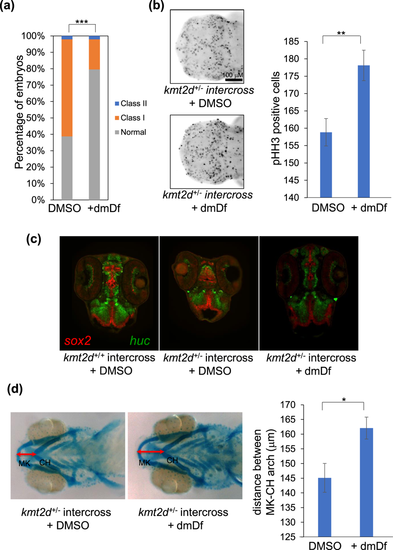
Treatment with dmDf rescues the defects of stable kmt2d mutant. kmt2d+/− adults were in-crossed and the embryos were then collected at 1 dpf, 2 dpf and 5 dpf to assess dmDf efficacy. (a) dmDf ameliorates significantly the CE defect in kmt2d+/− × kmt2d+/− progeny. (b) Treatment with dmDf increases significantly the brain cell proliferation of kmt2d+/− × kmt2d+/− progeny. (c) administration of dmDf appears to ameliorate the differentiation delay in kmt2d+/− × kmt2d+/− progeny. (d) dmDf rescues significantly the jaw defect in kmt2d+/− × kmt2d+/− progeny. *p < 0.05; **p < 0.01; ***p < 0.001. Error bars show SEM (standard error of the mean). |
|
ZFIN is incorporating published figure images and captions as part of an ongoing project. Figures from some publications have not yet been curated, or are not available for display because of copyright restrictions. PHENOTYPE:
|
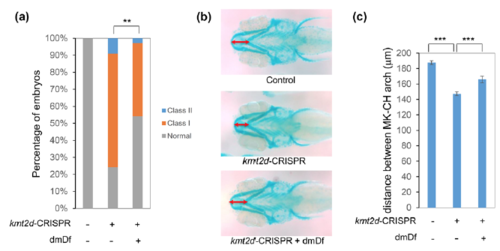
Treatment with dmDf rescues the developmental defects of kmt2d-CRISPR F0 mutants. (a) Similar to kmt2d-MO, kmt2d-CRISPR F0s also exhibit CE defects, and treatment with dmDf ameliorates the CE defects in kmt2d-CRISPR F0 embryos. (b) 5 dpf embryos were stained with Alcian blue to visualize jaw layout. kmt2d-CRISPR F0s exhibit the same change in jaw layout as kmt2d-MO-injected embryos, with a reduced distance between MK and CH cartilages. Treatment with dmDf ameliorates the jaw defects seen in kmt2d-CRISPR F0 embryos. (c) Quantitative measurement of the distance between MK and CH cartilages. ***: p< 0.001. Error bars show SEM (standard error of the mean). |