
FIGURE SUMMARY
- Title
-
A novel transgenic zebrafish line for red opsin expression in outer segments of photoreceptor cells
- Authors
- Crespo, C., Soroldoni, D., Knust, E.
- Source
- Full text @ Dev. Dyn.
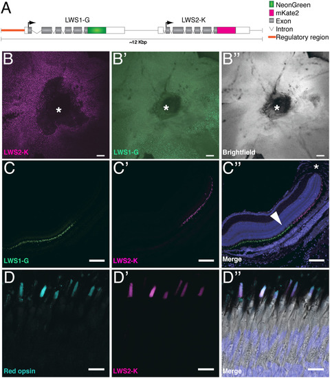
LWS1‐G and LWS2‐K are detected in outer segment of photoreceptor cells. A: Schematic illustration of Tg(LWS) (∼12 Kb, not to scale). Opn1lw1 and Opn1lw2 are C‐terminally fused to mNeonGreen and mKate2, respectively. The regulatory region (orange line) as defined by Tsujimura et al. (2010). The arrow depicts the orientation of the ORF. B–B″: Flat‐mounted retina of Tg(LWS) with LWS2‐K in magenta, LWS1‐G in green, and brightfield in gray. Asterisks outline the optic nerve of the retina. C–C″: Retinal section of an adult Tg(LWS) transgenic fish, ventral to the optic nerve (asterisk in C″). C″ shows the merge of C, C′, and DAPI staining (blue). Fluorescence of LWS1‐G is marked in green and LWS2‐K in magenta. Arrowhead in C″ highlights the outer nuclear layer. D–D″: Section of a Tg(LWS) adult retina showing staining with a red opsin antibody (cyan, D, D″) and fluorescence of LWS2‐K (magenta, D′, D″). D″ shows the merge of red opsin antibody staining (cyan), LWS2‐K fluorescence (magenta), DAPI staining (blue), and brightfield images. Scale bars B–C″ = 100 μm. Scale bars D–D″ = 10 μm.
|
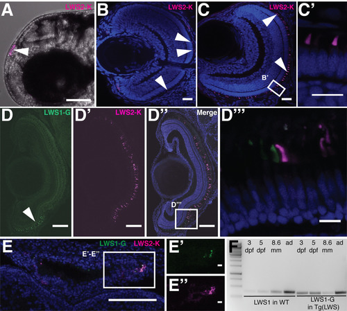
LWS1‐G and LWS2‐K protein expression during Zebrafish retinal development. A: LWS2‐K expression in the pineal gland (arrowhead) of a live Zebrafish embryo at 2 dpf. B–C: Retinal section of Tg(LWS) at 3 dpf with LWS2‐K in magenta and DAPI in blue. White arrowhead points to LWS2‐K in outer segment. D–D′: Retina of a Tg(LWS) animal of 8.6‐mm standard length, showing LWS1‐G in green and LWS2‐K in magenta. D″: Merge of D and D′ with DAPI in blue. D″′: Zoom in on boxed area in D″. E: Overview of a pineal gland of a Tg(LWS) larva at 8.6‐mm standard length with LWS2‐K in magenta, LWS1‐G in green, and DAPI in blue. E′–E″: Zoom in on boxed area in E. F: Reverse transcription polymerase chain reaction results for wild‐type (lane 2 to 5) and TG(LWS) (lane 6 to 9) littermates. cDNA was prepared from 3‐dpf, 5‐dpf, 8.6‐mm standard‐length larvae and adult retinas (ad). Wild‐type amplicons were obtained using opn1lw1 specific primers. TG(LWS) Polymerase chain reaction products were amplified using mNeonGreen specific primers. Lane 1: marker. Scale bar A = 1 mm. Scale bars B, C, D‐D″, E =100 μm. Scale bars D″′, E′, E″ = 10 μm. Scale bar C′ = 10 μm.
|
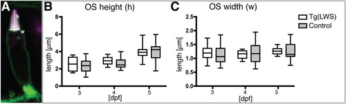
No obvious changes were detected in outer segment size of Tg(LWS) PRCs. A: Photoreceptor cell of Tg(LWS)/Tg(Ola.Actb:Hsa.HRAS‐EGFP)vu119 at 3 dpf showing mKate2 in magenta and green fluorescent protein at the plasma membrane in green. Outer segment height (h, dotted line) was calculated by measuring the distance from the midpoint at the base of the outer segment to the tip. Outer segment width (w, full line) was calculated by measuring the length of the OS base. B,C: Outer segment height (B) and width (C) were measured in both Tg(LWS)/ Tg(Ola.Actb:Hsa.HRAS‐EGFP)vu119 (white box) and Tg(Ola.Actb:Hsa.HRAS‐EGFP)vu119 stained with red opsin antibody (gray box with dots) at 3 dpf, 4 dpf, and 5 dpf. Measurements are represented in box plots with calculated minimum, 25th percentile, mean, 75th percentile, and maximum. The x‐axis shows the age of the embryos in days postfertilization, and the y‐axis shows the length measured in μm. For all time points and different transgenic lines, at least three independent samples were used and 20 measurements were carried out. Statistical significance was calculated by a one‐way ANOVA followed by Tukey's multiple comparison test. No statistically significant differences were observed.
|
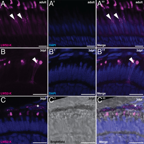
LWS2‐K signals in embryonic and adult retinas. Retinal sections of Tg(LWS) with LWS2‐K in magenta and DAPI in blue. A–B″: Vesicle‐like structures are present in the inner segment of LWS2‐K–positive cells. A–A″: Retinal sections of adult Tg(LWS). B–B″: Retinal sections of Tg(LWS) at 3 dpf. C–C″: Retinal sections of Tg(LWS) with brightfield image in gray scale. Asterisks indicate the LWS2‐K–positive signal in the retinal pigment epithelium, which is likely due to the pigment present in these cells. Scale bars A–C″ = 10 μm.
|
Acknowledgments
This image is the copyrighted work of the attributed author or publisher, and
ZFIN has permission only to display this image to its users.
Additional permissions should be obtained from the applicable author or publisher of the image.
Full text @ Dev. Dyn.