
- Title
-
The expression patterns of vestigial like family member 4 genes in zebrafish embryogenesis
- Authors
- Xue, C., Wang, H.H., Zhu, J., Zhou, J.
- Source
- Full text @ Gene Expr. Patterns
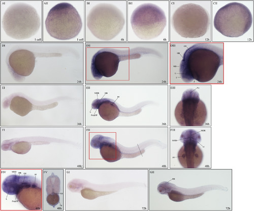
Expression of vgll4a in zebrafish embryos analyzed by WISH. Expression of vgll4a at one cell stage (AII), 6 hpf (BII), 12 hpf (CII), 24 hpf (DII-DIII), 36 hpf (EII-EIII), 48 hpf (FII-FV) and 72 hpf (GII). Embryos incubated with vgll4a sense probe are shown as negative controls (AI, BI, CI, DI, EI, FI, GI). Embryos are shown in lateral view with anterior to the left (DI-EII, FI, FII, FIV, GI, GII) or dorsal view with anterior to the top (EIII, FIII). Box areas in (DII) and (FII) was shown enlarged in (DIII) and (FIV) respectively. Dotted line in (FII) indicates approximate orientation of cross section image showed in (FV). T, telencephalon; E, eyes; MB, midbrain; HB, hindbrain; OV, otic vesicle; P, pharynx; L, lens; PF, pectoral fin; PA, pharyngeal arches; PP, pharyngeal pouches; Ve, ventricle; MHB, midbrain–hindbrain boundary; MDR midline of diencephalic roof; r1, rhombomere 1; Ed, endoderm; Ep, epidermis. EXPRESSION / LABELING:
|
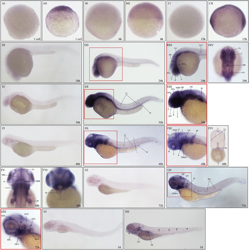
Expression of vgll4b in zebrafish embryos analyzed by WISH. Expression of vgll4b at one cell stage (AII), 6 hpf (BII), 12 hpf (CII), 24 hpf (DII-DIV), 36 hpf (EII-EIII), 48 hpf (FII-FVI), 72 hpf (GII-GIII) and 5 dpf (HII). Embryos incubated with vgll4b sense probe are shown as negative controls (AI, BI, CI, DI, EI, FI, GI, HI). Embryos are shown in lateral view with anterior to the left (DI-DIII, EI-FIII, GI-HII), dorsal view with anterior to the top (DIV, FV) or ventral view with anterior to the top (FVI). Box areas in (DII), (EII) and (FII) was shown enlarged in (DIII), (EIII) and (FIII) respectively. Dotted line in (FII) indicates approximate orientation of cross section image showed in (FIV). T, telencephalon; MTc, midbrain tectum; MTg, midbrain tegmentum; HB, hindbrain; LLP, lateral line primordium; P, pharynx; LL, lateral line; Pr, proctodeum; R, retina; L, lens; MHB, midbrain–hindbrain boundary; OfV, olfactory vesicle; OV, otic vesicle; PF, pectoral fin; N, neuromasts; Ep, epidermis; r, rhombomeres; r1, rhombomere 1; PP, pharyngeal pouches; PA, pharyngeal arches; Ed, endoderm; Ve, ventricle; H, heart; PLL, posterior lateral line; ION, infraorbital neuromasts; SON, supraorbital neuromast; OCN, occipital neuromasts; OLN, otic lateral neuromasts; MLN, middle line neuromasts; DTN, dorsal trunk neuromast; SB, swimming bladder. EXPRESSION / LABELING:
|
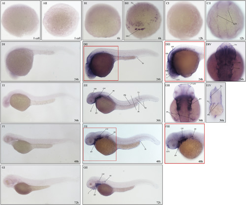
Expression of vgll4l in zebrafish embryos analyzed by WISH. Expression of vgll4l at one cell stage (AII), 6 hpf (BII), 12 hpf (CII), 24 hpf(DII-DIV), 36 hpf (EII-EIV), 48 hpf (FII-FIII) and 72 hpf (GII). Embryos incubated with vgll4l sense probe are shown as negative controls (AI, BI, CI, DI, EI, FI, GI). Embryos are shown in lateral view with anterior to the left (DI, DII, EI, EII, FI, FII, GI, GII) or dorsal view with anterior to the top (CI, CII, DIV, EIII). Box areas in (DII) and (FII) was shown enlarged in (DIII) and (FIII) respectively. Dotted line in (EII) indicates approximate orientation of cross section image showed in (EIV). Ep, epidermis; DFC, dorsal forerunner cells; EpN, epidermis at the border of the neural plate; Pr, proctodeum; OfV, olfactory vesicle; T, telencephalon; E, eyes; HB, hindbrain; OV, otic vesicle; Ve, ventricle; PP, pharyngeal pouches; P, pharynx; PF, pectoral fin; PD, pronephric duct; LL, lateral line; J, jaw. EXPRESSION / LABELING:
|

ZFIN is incorporating published figure images and captions as part of an ongoing project. Figures from some publications have not yet been curated, or are not available for display because of copyright restrictions. |
Reprinted from Gene expression patterns : GEP, 28, Xue, C., Wang, H.H., Zhu, J., Zhou, J., The expression patterns of vestigial like family member 4 genes in zebrafish embryogenesis, 34-41, Copyright (2018) with permission from Elsevier. Full text @ Gene Expr. Patterns