
- Title
-
Cytoplasmic polyadenylation-mediated translational control of maternal mRNAs directs maternal to zygotic transition
- Authors
- Winata, C.L., Łapiński, M., Pryszcz, L., Vaz, C., Bin Ismail, M.H., Nama, S., Hajan, H.S., Lee, S.G.P., Korzh, V., Sampath, P., Tanavde, V., Mathavan, S.
- Source
- Full text @ Development
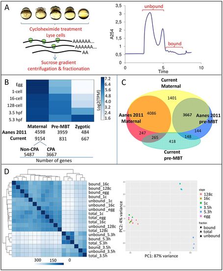
Polysome profile of the early embryo. (A) Polysome fractionation based on density gradient centrifugation. For each stage, two pooled fractions were obtained that contained polysome-unbound and polysome-bound mRNAs. (B) Gene expression clustering across developmental stages. Genes from Aanes et al. (2011) and the present dataset were clustered into maternal, pre-MBT and zygotic clusters. The maternal cluster was further subdivided based on pre-MBT increase in expression of transcripts that underwent CPA in the Aanes dataset that was attributable to oligo d(T) selection bias. (C) Venn diagram showing overlap between maternal and pre-MBT clusters from both datasets. Notice the large overlap between the Aanes pre-MBT and present maternal cluster. (D) Clustering heat-map and principal component analysis of total and polysome-fractionated samples based on the expression of annotated genes (Ensembl), showing a major division between early and late (MBT and post-MBT) developmental stages. Clustering was performed based on the abundance of transcript level to compute the distances between samples (different color intensity). |
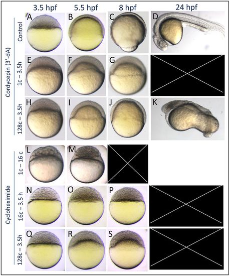
Epiboly defects caused by 3′dA and CHX treatments. (A-D) Untreated control embryos. (E-G) Embryos that were treated with 3′-dA from the one-cell stage to 3.5 hpf undergo developmental arrest and cytolysis before 24 hpf (n=40). (H-K) 3′-dA treatment from the 128-cell stage to 3.5 hpf caused a delay of epiboly and gross patterning defects (n=30). (L,M) Translation inhibition by CHX treatment that was initiated at the one-cell stage affected early development and caused early lethality (n=20), whereas CHX treatment initiated at the 16-cell stage (N-P) resulted in developmental arrest at the oblong stage (n=61). Notice the larger cells at 3.5 hpf, suggesting defects in cell division. (Q-S) Treatment that was initiated at the 128-cell stage resulted in epiboly arrest followed by mortality (n=66). |
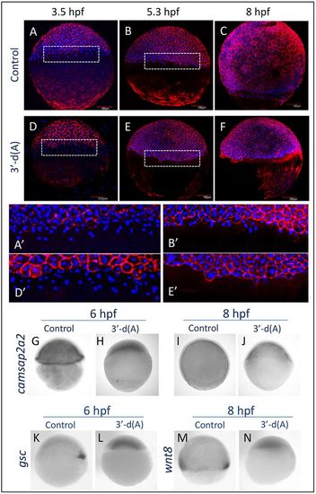
Phenotype caused by CPA inhibition. (A-C) At 3.5 and 5.3 hpf, the external yolk syncytial layer (e-YSL) could be observed in control embryos. (D-F) Embryos treated with 3′-dA beginning at the one-cell stage to 3.5 hpf still had visible e-YSL at 3.5 hpf, but this structure disappeared by 5.3 hpf, and epiboly did not proceed further (n=5). (G-J) Whole-mount in situ hybridization with the YSL marker camsap2a2 confirmed the absence of e-YSL up to 8 hpf (n=8). Other gastrulation markers, including dorsal shield (K-L; n=8) and mesendodermal cells expressing wnt8 (M,N; n=8), were also absent. EXPRESSION / LABELING:
PHENOTYPE:
|