
- Title
-
Identification of an Evolutionarily Conserved Ankyrin Domain-Containing Protein, Caiap, Which Regulates Inflammasome-Dependent Resistance to Bacterial Infection
- Authors
- Tyrkalska, S.D., Candel, S., Pérez-Oliva, A.B., Valera, A., Alcaraz-Pérez, F., García-Moreno, D., Cayuela, M.L., Mulero, V.
- Source
- Full text @ Front Immunol
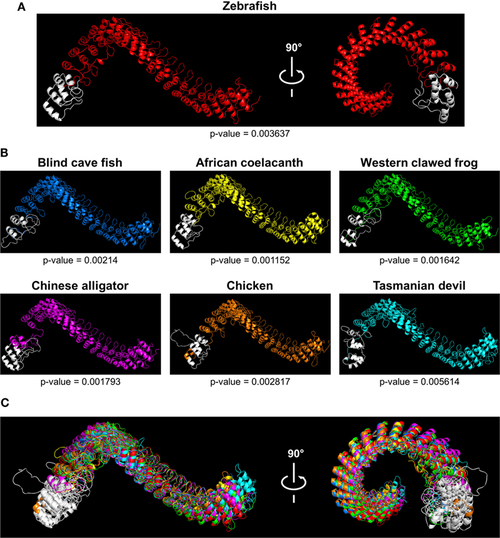
Tridimensional models of Caiap structures in different species. (A,B) 3D models showing Caiap proteins from zebrafish (Danio rerio) (A), and blind cave fish (Astyanax fasciatus mexicanus), western clawed frog (Xenopus tropicalis), African coelacanth (Latimeria chalumnae), Chinese alligator (Alligator sinensis), chicken (Gallus gallus), and Tasmanian devil (Sarcophilus harrisii) (B) with corresponding accuracies. (C) The 3D Caiap models from all species shown in (A,B) were superimposed. The CARD domains are shown in white. |

ZFIN is incorporating published figure images and captions as part of an ongoing project. Figures from some publications have not yet been curated, or are not available for display because of copyright restrictions. PHENOTYPE:
|
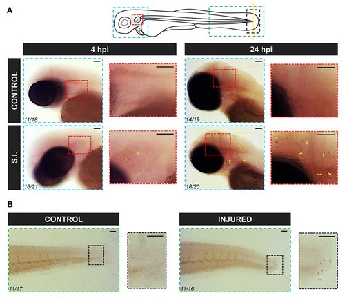
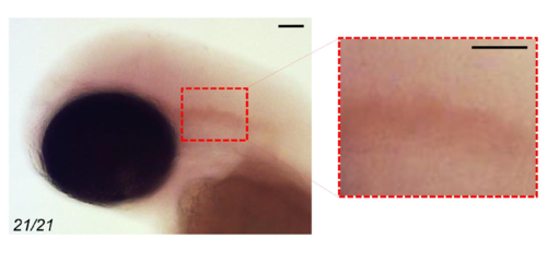
Zebrafish caiap is induced in discrete cells at the infection site. Zebrafish 2 dpf larvae were infected with ST in the otic vesicle (A) or tail wounded (B). At the indicated times, WISH was performed using antisense probes to the caiap gene. Note the presence of caiap+ cells at both the infection and injured sites (arrowheads). The areas shown are indicated in the larval scheme with boxes of different colors. Numbers in pictures represent the animals with the shown phenotype per total analyzed animals. S.I., ST infection. Scale bar: 0.5 mm. EXPRESSION / LABELING:
PHENOTYPE:
|
|
ZFIN is incorporating published figure images and captions as part of an ongoing project. Figures from some publications have not yet been curated, or are not available for display because of copyright restrictions. PHENOTYPE:
|
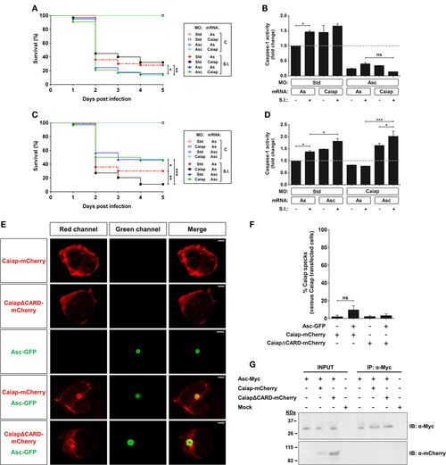
Asc is required for the Caiap-mediated resistance to S. Typhimurium. (A–D) Zebrafish one-cell embryos were injected with standard control (Std) (A–D), Asc (A,B) or Caiap (C,D) MOs or in combination with antisense (As) (A–D), Caiap (A,B) or Asc (C,D) mRNAs, infected at 2 dpf and survival (A,C) and caspase-1 activity (B,D) determined as described in Figures 5A,B, respectively. S.I., ST infection. (E–G) HEK293T cells were transfected with zebrafish Caiap-mCherry or CaiapΔCARD-mCherry in the presence or absence of zebrafish Asc-Myc, fixed at 48 h post-transfection, immunostained and imaged using a laser confocal microscope (E,F) or lysed, immunoprecipitated with ANTI-Myc Affinity Gel, and probed with antibodies to Myc and mCherry (G). Representative views of maximum-intensity projection images of HEK293T cells are shown in (E), quantitation of the percentage of Caiap specks in relation to the total number of Caiap transfected cells is shown in (F) and representative blots are shown in (G). The sample size for each treatment is 300 for (A,C), 30 for (B,D), and 50 for (F). Scale bar, 5 µm; ns, not significant; *p < 0.05; **p < 0.01; ***p < 0.001. PHENOTYPE:
|
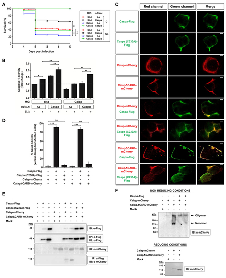
Caspa is required for the Caiap-mediated resistance to S. Typhimurium. (A,B) Zebrafish one-cell embryos were injected with standard control (Std) or Caiap MOs in combination with antisense (As) or Caspa mRNAs. At 2 dpf, embryos were infected with ST and survival (A) and caspase-1 activity (B) were determined as described in Figures 5A,B, respectively. S.I., ST infection. (C–F) HEK293T cells were transfected with zebrafish Caiap-mCherry or CaiapΔCARD-mCherry in the presence or absence of zebrafish wild-type FLAG-Caspa or catalytic inactive FLAG-Caspa (C230A), fixed at 48 h post-transfection and imaged using a laser confocal microscope (C,D), or lysed and immunoprecipitated with ANTI-FLAG M2 Affinity Gel and probed with antibodies to FLAG and mCherry (E) or lysed and resolved under non-reducing and reducing conditions (F). Representative views of maximum-intensity projection images of HEK293T cells are shown in (C), quantitation of the percentage of Caiap specks in relation to the total number of Caiap transfected cells is shown in (D) and representative blots are shown in (E,F). The sample size for each treatment is 300 for (A), 30 for (B), and 50 for (C). Scale bar, 5 µm; not significant; *p < 0.05; **p < 0.01; ***p < 0.001. |
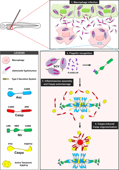
Proposed model illustrating the stabilization of catalytic active Caspa tetramers by Caiap. 1. Macrophages are recruited to the infection site where they are infected by S. Typhimurium (ST), which is contained within the Salmonella-contained vesicle (SCV). 2. Flagellin is inadvertently injected into the cytosol though the type 3 secretion system of ST where it is recognized by a Nlr. 3. The inflammasome is assembled via formation of Asc specks. 4. Active Caspa interacts with Caiap via its ANK domains and induces its self-oligomerization through CARD/CARD allowing Caspa stabilization in high molecular weight complexes upon its prodomain release. |
Control of WISH results shown in Figure 4. Zebrafish 2 dpf larvae were infected with ST in the otic vesicle. WISH was performed at 4 hpi using sense probes to the caiap gene. Note that no positive cells were observed at the infection site. The area shown is indicated in the larval scheme with boxes of different colors in Figure 4. Numbers in pictures represent the animals with the shown phenotype per total analyzed animals. Scale bar: 0.5 mm. |
|
ZFIN is incorporating published figure images and captions as part of an ongoing project. Figures from some publications have not yet been curated, or are not available for display because of copyright restrictions. PHENOTYPE:
|