
- Title
-
Retrotransposons Mimic Germ Plasm Determinants to Promote Transgenerational Inheritance
- Authors
- Tiwari, B., Kurtz, P., Jones, A.E., Wylie, A., Amatruda, J.F., Boggupalli, D.P., Gonsalvez, G.B., Abrams, J.M.
- Source
- Full text @ Curr. Biol.
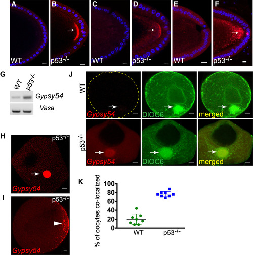
Germ Plasm Localization Is Conserved in Invertebrates and Vertebrates (A–F) Patterns of other retrotransposons in the fly oocyte were profiled by in situ hybridization. Examples of Tahre (A and B), Burdock (C and D), and HeT-A (E and F) on WT (A, C, and E) and p53−/− (Β, D, and F) oocytes are shown. DAPI counterstain is indicated in blue. Like Tahre, Burdock, and HeT-A RNAs also localized to the germ plasm region (arrows). (G–K) Gypsy54 localizes to the germ plasm of zebrafish oocytes. (G) RT-PCR analysis of Gypsy54 and vasa (loading control) from WT and p53−/− zebrafish ovaries. (H)–(J) show in situ hybridizations for Gypsy54 RNA (red) on WT and p53−/− oocytes. Arrows point to the Balbiani body, the presumptive germ plasm of stage I oocytes (H). The arrowhead in (I) indicates the germ plasm at the cortical region of stage II oocytes. DiOC6 (green) was used to label the Balbiani body (J, arrows) in p53−/− and WT oocytes. Images are single optical sections. The scatterplot in (K) shows percentages of DiOC6-stained Balbiani bodies that were also positive for Gypsy54 RNA. Scale bars, 10 μm. See also Figure S4 and STAR Methods. |
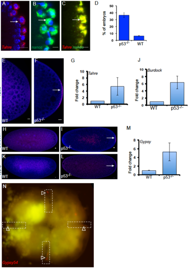
Figure S4. Tahre elements from the oocyte germ plasm persist in the embryonic germline. Related to Figures 3 and 4. Confocal sections at the cortex of p53-/- embryos labeled with Tahre (A), nanos (B), and co-localization of Tahre with nanos (C), (indicated by arrows) showing that similar to nanos, Tahre transcripts remain highly associated to the pole cells. Embryos 0-3 hr produced from either p53-/- females crossed with WT males or from WT females mated to p53-/- males were scored for the presence of Tahre FISH signal at the pole plasm (D). The maternal effect seen in these reciprocal crosses verifies that Tahre transcripts present in the presumptive germ cells were maternally derived. Tahre, Burdock and Gypsy retroelements are transmitted to the embryonic pole plasm. Confocal images of RNA in situ hybridization of Tahre (E-G), Burdock (H-J) and Gypsy (K-M) on WT and p53-/- (0-2 h) embryos. FISH signal at the pole plasm is shown by arrow. Gypsy transcripts were also detected in the pole plasm of early embryos and could occur below detection limits in the oocyte germ plasm [S5, S6]. Note FISH signals are at the pole plasm in p53-/- embryos. Bar graph quantifies the fold change of the FISH positive signal at the posterior pole of p53-/- embryos relative to WT (0-2 h) from three biological replicates, n=50. DAPI counterstain (blue). Gypsy54 mRNAs localize to the cleavage plane of the Zebrafish embryos. Gypsy54 RNA in situ hybridization on the 4 cell staged p53-/- Zebrafish embryos (N). Dotted lines denote the cleavage plane and arrowheads indicate Gypsy54 signal at the presumptive germline. Scale bars: 10 μm. Error bars represent ±SD. |