
- Title
-
Wolf-Hirschhorn Syndrome Candidate 1 (whsc1) Functions as a Tumor Suppressor by Governing Cell Differentiation
- Authors
- Yu, C., Yao, X., Zhao, L., Wang, P., Zhang, Q., Zhao, C., Yao, S., Wei, Y.
- Source
- Full text @ Neoplasia
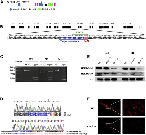
Generation of whsc1 mutant zebrafish with Crispr/Cas9 technology. (A) A diagram showing the structure of WHSC1 protein. (B) design of sgRNA against whsc1 gene. This sgRNA site targets the 15th exon. Target sequence was shown with blue letters, and PAM region was shown in red. Green box marked the reorganization site of BSTX1 enzyme that lies within sgRNA target site, loss of which indicated the presence of indels. (C) Restriction enzyme analysis of the presence of indel mutations in two independent mutant lines. (D) Sanger sequencing results of the whsc1 mutant lines. Arrows indicated the locations of indels. (E) WB analysis of the levels of H3K36Me2 and H3K36Me3 in whsc1 wild-type and mutant embryos. (F) Immunofluorescence analysis of H3K36Me2 level in whsc1 wild-type and mutant embryos. Photos of wildytpe and whsc1 mutant embryos were taken under same exposure time. |
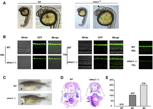
WHS related phenotypes in whsc1−/− zebrafish. (A) Brain morphology in whsc1−/− and wild-type embryos. Arrow heads point to the diencephalic ventricle (homologue of mammalian third ventricle). (B) Motor neuron development in whsc1−/− and wild-type embryos. Note that the number of motor neurons in whsc1−/− mutant larva was significantly less than that in wild-type ones. (C) whsc1−/− adult zebrafish showed heart enlargement. (D) Tissue section analysis of the heart of wild-type or mutant adult fishes. Arrows point to the hearts. (E) Statistics of heart enlargement in these two mutation lines. PHENOTYPE:
|
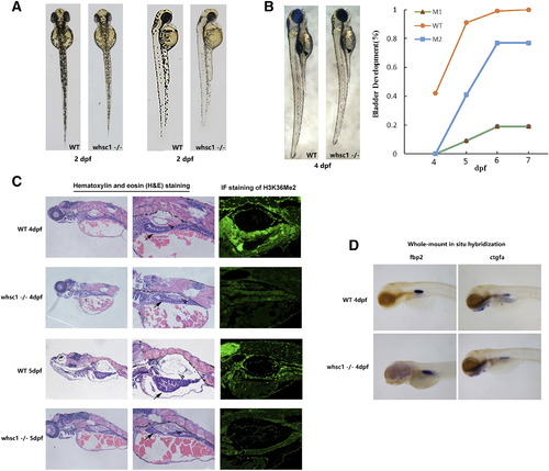
whsc1−/− zebrafishes had developmental defects in swim bladder. (A) In the early development, compared to age-matched wild-type embryos, whsc1−/− embryos showed reduced body length and less pigmentation. (B) The left part shows that swim bladder inflation defects in whsc1−/− zebrafish. Arrow heads point to the swim bladders. Note that wild-type larva had already inflation, while whsc1−/− larva had not, suggesting their swim bladders were either developed abnormally or not functional. And the right part is the statistics of inflation of swim bladder in whsc1−/− zebrafish and wild-type zebrafish. (C) HE staining and immunofluorescence analysis of the swim bladder in whsc1−/− zebrafish and wild-type zebrafish at 4 dpf and 5 dpf. Arrow heads pointed to the bladder and arrows pointed to the gut. In the HE staining of zebrafish larvae at 4 and 5 dpf (left and middle panels), bladders in wild-type larvae had formed a three layer structure that was filled with air. However, bladders in whsc1 mutants were still cell masses and failed to develop three layers. Immunofluorescence staining of the H3K36me2 revealed that whsc1−/− larvae expressed significantly lower level of H3K36me2 than wild-type larvae did. (D) WISH analysis of swim bladder markers fbp2 and ctgfa in whsc1−/− zebrafish and wild-type zebrafish at 4 dpf. |
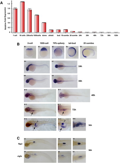
whsc1 expression pattern during early embryonic development. (A) Quantitative PCR analysis of whsc1 mRNA levels in embryos at various developmental stages. Relative ratio (fold changes) obtained from 1-cell stage embryos was set to 1. (B) Whole mount in situ hybridization analysis of whsc1 mRNA expression pattern in embryos at various developmental stages. B1-B2 were lateral view; B3-B4 were lateral view with dorsal to the right; B7, B9, B11 and B16 were dorsal view with head to the left; the rest were lateral view with head to the left. Note that whsc1 was expressed specifically in the swim bladder and gut (B14–15). Arrowheads point to the bladder and arrows point to the gut. (C) WISH analysis of swim bladder markers fbp2 and ctgfa. C1–2 and C4–5 were shown in lateral view with head to the left; C3 and C6 were shown in dorsal view with head to the left. |
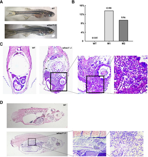
whsc1 mutant zebrafish developed swim bladder lesions. (A) One-month-old whsc1−/− zebrafishes developed skin lesions. Arrow pointed to the lesion. (B) The Statistics of wild-type and whsc1 mutant fishes with lesions. (C) HE staining of swim bladder in one-month-old wild-type and whsc1 mutant zebrafishes (cross-section). Tissues were serially sectioned from head to tail, and sections containing swim bladders (wild-type fishes) or skin lesions (mutant fishes) were HE stained for analysis. (D) HE analysis of swim bladder in three-month-old zebrafish (vertical section). Note that wild-type fish had normal and functional swim bladder, while whsc1 mutant fishes had no obvious swim bladder. The position of swim bladder in whsc1 mutant was occupied with cell masses that seemed to have no cell polarity. PHENOTYPE:
|
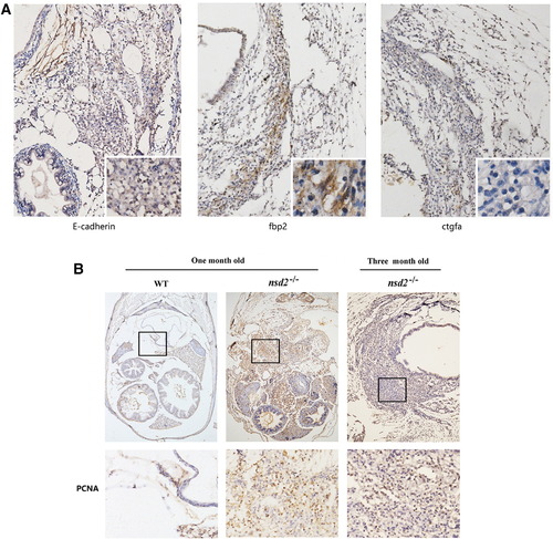
IHC staining of the zebrafish swim bladder tumor. (A) IHC staining of E-cadherin, fbp2 and ctgfa in tumor tissues of whsc1 mutant zebrafishes. (B) IHC staining of the PCNA expression in normal zebrafish swim bladder and tumor tissues of whsc1 mutant fishes. EXPRESSION / LABELING:
PHENOTYPE:
|

ZFIN is incorporating published figure images and captions as part of an ongoing project. Figures from some publications have not yet been curated, or are not available for display because of copyright restrictions. |
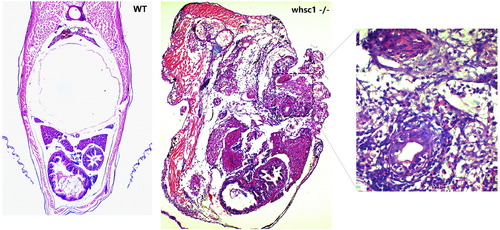
HE analysis of pathological tissue in one-month-old zebrafish (cross-section). |