
- Title
-
Bioenergetic status modulates motor neuron vulnerability and pathogenesis in a zebrafish model of spinal muscular atrophy
- Authors
- Boyd, P.J., Tu, W.Y., Shorrock, H.K., Groen, E.J.N., Carter, R.N., Powis, R.A., Thomson, S.R., Thomson, D., Graham, L.C., Motyl, A.A.L., Wishart, T.M., Highley, J.R., Morton, N.M., Becker, T., Becker, C.G., Heath, P.R., Gillingwater, T.H.
- Source
- Full text @ PLoS Genet.

ZFIN is incorporating published figure images and captions as part of an ongoing project. Figures from some publications have not yet been curated, or are not available for display because of copyright restrictions. |

Overexpression of necdin ameliorates the motor axon outgrowth phenotype in smn morphant zebrafish. (A) Western blotting of cytochrome C, an electron transport chain protein showed an increase in NDN overexpressing embryos suggesting an increase in mitochondrial biogenesis. (B) Cyt C protein levels were quantified relative to a loading control. (C) Representative confocal micrographs of motor neuron axons exiting the spinal cord in control (top), smn morphant (middle) and smn morphant over-expressing Ndn (bottom) Tg(hb9:GFP) zebrafish embryos. Note the presence of the axonal outgrowth phenotype associated with smn knockdown (arrow heads) is reduced in the Ndn expressing animals. Scale bars = 50 μM. (D) Bar chart (mean & s.e.m.) showing a significant increase in the number of normal MNs, and a concomitant significant decrease in the number of severely affected MNs, in co-injected smn MO and Ndn mRNA embryos compared to single smn MO injected embryos at 30 hpf. Unpaired two-tailed student t-tests; * p<0.05, ** p<0.01 *** p<0.001. N = 20 embryos per experimental group. |

Pgk1 expression is pathologically relevant in mouse and zebrafish models of SMA. (A) Expression of PGK1 protein in the spinal cord, skeletal muscle, sciatic nerve and heart of late-symptomatic P8 SMA mice. Protein levels were quantified and normalized to an appropriate loading control. (B) Bar chart (mean & s.e.m.) showing a significant reduction in PGK1 protein levels in SMA mouse spinal cord and sciatic nerve. N = 6 SPC per genotype. N = 3 muscle per genotype. N = 7 sciatic nerves per genotype. N = 3 hearts per genotype (C) Knockdown of Pgk1 in zebrafish induced an axonal outgrowth phenotype (middle panel arrow) similar to smn knockdown (arrow bottom panel) and also produced swellings in the tips of outgrowing axons indicative of axonal transport deficiencies. Scale bars = 50 μM (D) Quantification of axonal outgrowths showed a significant increase in truncated motor axons in pgk1 and smn morphants compared to controls. (E) Efficiency of pgk1 knockdown in embryos was shown by western blot embryos normalized to an appropriate loading control (N = 3 per group, batches of 30 pooled zebrafish embryos per lane). N = 20 embryos per group. Unpaired two-tailed students t-test * p<0.05, ** p<0.01 *** p<0.001 **** p<0.0001. PHENOTYPE:
|
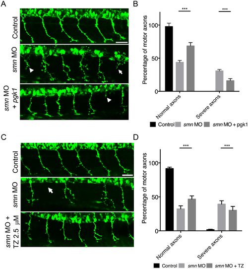
Overexpression or pharmacological activation of pgk1 rescues motor neuron phenotypes in smn morphant zebrafish. (A) Representative confocal micrographs of primary motor neuron axons exiting the spinal cord in control (top), smn morphant (middle) and smn morphant over-expressing pgk1 (bottom) Tg(hb9:GFP) zebrafish embryos. Note the presence of an axonal outgrowth/branching phenotype associated with smn knockdown (arrow heads) that is reduced in the pgk1 over-expressing animals. Scale bars = 50 μM. (B) Overexpression of Pgk1 in smn morphant zebrafish at 30 hpf led to a significant increase in normal motor axons and significant decrease in severe axonal outgrowth phenotypes compared to single smn MO injected embryos. (C) Representative confocal micrographs of motor neuron axons exiting the spinal cord in control (top), smn morphant (middle) and smn morphant treated with 2.5 μM terazosin (bottom) Tg(hb9:GFP) zebrafish embryos. Note how the presence of the axonal outgrowth/branching phenotype associated with smn knockdown (arrow heads) is reduced in the terazosin-treated animals. (D) Bar chart (mean & s.e.m) showing activation of Pgk1 by treatment with 2.5 μM terazosin in smn morphant zebrafish at 30 hpf led to a significant increase in normal motor axons and significant decrease in severe axonal outgrowth phenotypes compared to untreated smn MO injected embryos. Unpaired two-tailed student t-tests * p<0.05, ** p<0.01 *** p<0.001. n = 20 embryos per group. |
Knockdown of Smn via morpholino efficiency. (A) Efficiency of Smn knockdown as determined by western blot in 48hpf zebrafish. (B) knockdown was quantified and normalized to CoxIV loading control (N = 3 per group, batches of 30 pooled zebrafish embryos per lane). Bar chart (mean & s.e.m). Unpaired two-tailed student t-test * P<0.05. |
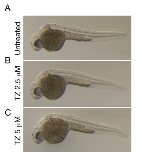
Treatment with terazosin does not cause any morphological or development defects in zebrafish embryos. Representative bright field of images 28 hpf zebrafish treated with terazosin. (A) Control untreated zebrafish. (B) Treatment of 2.5 μM Terazosin and (C) Treatment of 5 μM Terazosin (TZ) from 6 hpf does not lead to any morphological or developmental delay compared to untreated zebrafish. |