
- Title
-
Oncogenic role of rab escort protein 1 through EGFR and STAT3 pathway
- Authors
- Yun, U.J., Sung, J.Y., Park, S.Y., Ye, S.K., Shim, J., Lee, J.S., Hibi, M., Bae, Y.K., Kim, Y.N.
- Source
- Full text @ Cell Death Dis.
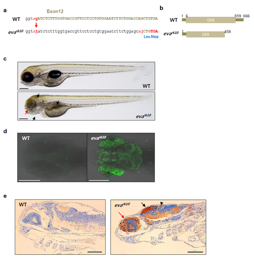
(a and b) The G to A substitution in splicing acceptor site of exon 12 in REP1 genes induced zebrafish evanescence (evark10) mutant having abnormal stop codon by aberrant splicing. (b) evark10 mutant protein lacks the C-terminal GDI domain of the wild-type REP1 protein of zebrafish. (c) Morphological phenotypes of WT and evark10 mutant embryos were examined with dissecting microscope at 5 day post fertilization (dpf). Arrows indicate abnormal morphology of head, pharyngeal arch, swim bladder (black), and eye (red). (d) Apoptosis was visualized by TUNEL assay in WT and evark10 mutant embryos at 5 dpf. (e) Paraffin-embedded zebrafish tissues were subjected to immunohistochemistry using anti-active caspase3 antibody. Eye: red arrow; Tectum: black arrow; Cerebellum: arrowhead Magnification: x50; scale bar = 100 µm. Similar results were observed in two independent experiments. PHENOTYPE:
|
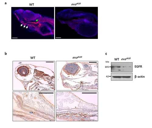
(a) Zebrafish frozen tissue sections were subjected to immunofluorescence assay using anti-EGFR antibody. Pharyngeal arch: white arrows; Esophagus: green arrow; EGFR: red; Nucleus; blue. Magnification: x50, Scale bar = 100 µm. (b) Paraffin-embedded zebrafish tissues were subjected to immunohistochemistry using anti-EGFR antibody. Scale bar = 100 µm. (c) Cell lysates from WT and evark10 mutant embryos at 5dpf were processed for immunoblot analysis using anti-EGFR and β-actin antibodies. β-actin was used as a loading control. Similar results were observed in two independent experiments. |