
- Title
-
ATP6V1H Deficiency Impairs Bone Development through Activation of MMP9 and MMP13
- Authors
- Zhang, Y., Huang, H., Zhao, G., Yokoyama, T., Vega, H., Huang, Y., Sood, R., Bishop, K., Maduro, V., Accardi, J., Toro, C., Boerkoel, C.F., Lyons, K., Gahl, W.A., Duan, X., Malicdan, M.C., Lin, S.
- Source
- Full text @ PLoS Genet.
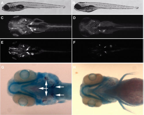
Bone but not cartilage cells are defective in atp6v1h-deficient zebrafish. (A-B) shows an image of a 6 days post fertilization (dpf) wild type and mutant embryo with relatively normal gross morphology. (C-F) Day 6 and 8 wild type and mutant embryos showing reduction in bone detected by calcein staining (ventral view, bright fluorescent stains indicate calcified bone). In (G-H), double staining for cartilage and bone demonstrate reduction of mineralized bone (dark purple, arrows) but not cartilage (blue). A, C, E and G are wild type. B, D, F, and H are mutant. |
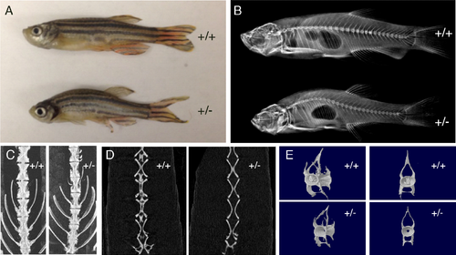
Micro-computed tomography scan of adult zebrafish. Adult zebrafish heterozygous for atp6V1h mutation (+/-) show curved body, compared to the wild type (+/+) sibling (A). X-ray of the same +/- fish shows curved spine compared to the +/+ sibling (B). Vertebra of +/- fish has bristlier surface and smaller size (C). Vertebra of +/- fish lacks calcified structure in the centrum cavity (D). Individual vertebra of +/- fish shows bristlier surface and smaller size with a hollow centrum (E) (n = 3 each). PHENOTYPE:
|

Induction of mmp9/mmp13 and rescue of atp6v1h-deficient phenotype. In (A), qPCR analysis of designated marker genes showing significant induction of mmp9 and mmp13 mRNA expression in atp6v1h mutant. Analysis of osteoclast cells by RNA whole mount in situ hybridization. atp6v1h mutant embryos showing significant increases of mmp9 and mmp13a signals in embryonic head skeleton (e/f/i/j wild type, g/h/k/l mutant), but osteoclast marker rank pattern is not changed (a/b wild type, c/d mutant) (B). In (C), analysis of Mmp9/Mmp13 expression in mouse osteoclast Raw264.7 cells after knockdown of Atp6v1h by siRNA is shown. When Atp6v1h expression was knocked down by siRNA (a), the expression of Mmp9 (b) and Mmp13 (c) were induced. Similarly, the induction of Mmp9 (d) and Mmp13 (e) was also observed in RANKL-induced differentiated mouse osteoclast Raw264.7 cells. In (D), MMP inhibition rescues the bone phenotype in Atp16V1h mutant fish as detected by calcein staining; a, wild type, 0.5% DMSO; b, mutant, 0.5% DMSO; c, mutant treated with 200uM MMP9 inhibitor II in 0.5% DMSO; d, mutant treated with 20uM MMP13 inhibitor in 0.5% DMSO; e, mutant treated with 50uM MMP9/13 inhibitor I in 0.5% DMSO. Images are ventral views of 6 dpf embryos. |
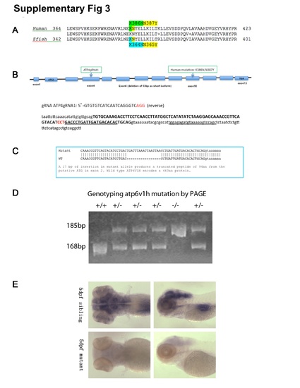
Generation and Genotyping of atp6v1h zebrafish mutants. Protein alignment of ATP6V1H in human (NP_057025.2) and zebrafish (NP_775377.1) shows high homology (~85%). More importantly, the region where the mutation is located is highly conserved (A). Using CRISPR/Cas9, guide RNA (gRNA) targeting exon 4 of zebrafish atp6v1h was co-injected with Cas9 mRNA into zebrafish embryos and detected by T7 endonuclease digestion. Sequence of gRNA and target is shown (B). Several founders with indels were screened for germline transmission, and the one with 17bp insertion was used throughout the study; The 17bp insertion is predicted to produce a termination codon 94 amino acids after initiation of start codon (C). Genotyping done by polyacrylamide gel electrophoresis (PAGE) analysis demonstrating a 168 bp band for wild type allele, and 185 bp band for mutant allele; representative results for wild type (+/+), heterozygous (+/-), and homozygous (-/-) embryos are shown (D). Using probes specific for atp6v1h, RNA in situ hybridization analysis of atp6v1h showed expression in the head region in wild type embryos, while the expression is nearly absent in homozygous atp6v1h embryos, suggesting that the 17bp insertion may lead to nonsense-mediated decay (NMD) of mRNA and result in loss-of-function of this gene (E). |
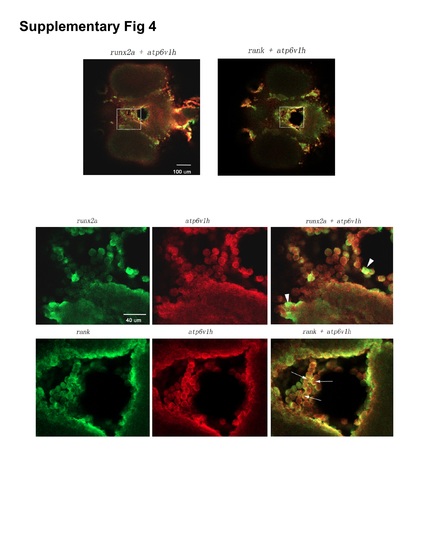
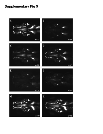
Co-expression analysis of atp6v1h with osteoclast or osteoblast markers. Double fluorescence RNA whole mount in situ hybridization with atp6v1h and rank or runx2a probes were performed on 60 hpf embryos and imaged by confocal microscopy. Upper panels are ventral views of merged lower magnification images of runx2a and atp6v1h or rank and atp6v1h, in which the boxes indicate the regions shown in the lower panels of higher magnification. The lower panels are images of individual colors (green fluorescence for probes of runx2a or rank, red fluorescence for atp6v1h) and merged color (runx2a + atp6v1h, rank+ atp6v1h). Arrowheads point to cells that are mainly positive for runx2a and arrows point to cells that appear positive both for rank and atp6v1h (co-expression: yellow fluorescence). EXPRESSION / LABELING:
|
Rescue of atp6v1h mutant zebrafish. Bone staining of uninjected wild type and atp6v1h -/- embryos; loss of bone mineralization is seen in atp6v1h mutant embryos (A and B). atp6v1h mutant embryos at single-cell stage injected with wild type mRNA (C, 200 pg; D, 300 pg) were stained at 5dpf; representative figures demonstrate the rescue of bone phenotype, seen as increase in bone mineralization. atp6v1h mutant embryos injected with mutant mRNA containing the same K386N and N387Y mutations found in humans (E, 200 pg; F, 300 pg) at single-cell stage and stained at 5 dpf; representative figures show the absence of bone staining, indicating that the mutations create a non-functional gene. Wild type and mutant mRNA were injected into wild type embryos at single-cell stage and stained at 5 dpf (G and H); representative figures show no alteration in morphology and bone staining, suggesting the absence of gain-of-function resulting from overexpression of either wild type or mutant mRNA. |
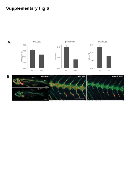
Micro-CT analysis of heterozygous adult bone phenotype. A. Micro-CT data of the first to fifth caudal vertebrae. Wild type sibling +/+. Heterozygous +/-. BMD, BV and BS designate bone mineral density, bone volume and bone surface (n = 3). B. Micro-CT images of bone in 8-month-old male adult wild type zebrafish and heterozygous allele of retrovirus insertion mutant, atp6v1hhi923+/- (n = 5 each). |
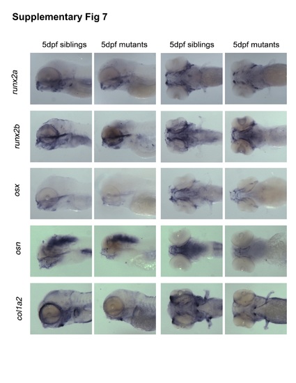
RNA in situ hybridization analysis of osteoblast and osteoclast gene markers. In situ hybridization markers of osteoblast development (runx2a, runx2b, osx, osn, col1a2) show slight reduction between 5dpf siblings (wild type) or mutant (-/-). |
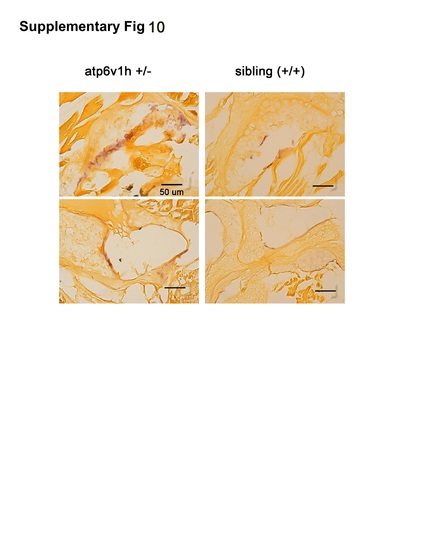
Analysis of osteoclast by TRAP in adult zebrafish. Adult zebrafish (10 month old) of +/- for atp6v1h (line atp6v1hla439) and wild type sibling were fixed, sectioned and stained by TRAP. Atp6v1hla439+/- fish appear to have more TRAP staining compared to their sibling (n = 3, two images are shown here for wild type and +/- mutant). Scale bar: 50 um. |

ZFIN is incorporating published figure images and captions as part of an ongoing project. Figures from some publications have not yet been curated, or are not available for display because of copyright restrictions. PHENOTYPE:
|