
- Title
-
GUCA1A mutation causes maculopathy in a five-generation family with a wide spectrum of severity
- Authors
- Chen, X., Sheng, X., Zhuang, W., Sun, X., Liu, G., Shi, X., Huang, G., Mei, Y., Li, Y., Pan, X., Liu, Y., Li, Z., Zhao, Q., Yan, B., Zhao, C.
- Source
- Full text @ Genet. Med.
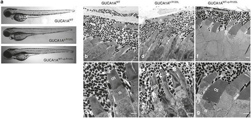
Morphological changes caused by GUCA1A p.R120L. (a) Light microscopy indicates no evident morphological changes in eyes of zebrafish injected with GUCA1AWT, or GUCA1Ap.R120L, or GUCA1AWT+p.R120L 4 days postfertilization (dpf). (b–e) Transmission electron microscopy of the retina of zebrafish 11 dpf injected with GUCA1AWT or GUCA1Ap.R120L. Photoreceptors are regularly lined and shaped in the GUCA1AWT-injected group (b, d). Thickness of the RPE; shrinking, twisty, and caducous photoreceptor outer segments; and choriocapillary disruptions are found in the GUCA1Ap.R120L-injected group (c, e). ▵ represents the RPE nucleus. Scale bar = 1 μm. |
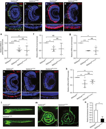
Impairments in photoreceptors, the retinal pigment epithelium (RPE), and ocular vasculature induced by GUCA1A p.R120L in zebrafish. (a–d) Immunofluorescent staining of rhodopsin (RHO) (red; a,b), peanut agglutinin (PNA) lectin (green; a,b), and Zpr-1 (red; c,d). RHO and PNA expressions were significantly reduced in zebrafish overexpressing GUCA1Ap.R120L (a,b). Zpr-1 expression was only slightly reduced in the GUCA1Ap.R120L-injected group (d) when compared with GUCA1AWT-injected zebrafish (c). (e–g) Relative messenger RNA (mRNA) levels of photoreceptor-, cone-, and rod-specific transcripts in GUCA1Ap.R120L- and GUCA1AWT+p.R120L-injected zebrafish compared with the GUCA1AWT-injected group. (h–j) Robust Zpr-2 expression was detected in retinal frozen sections of 4-dpf larvae injected with GUCA1AWT (h), whereas Zpr-2 expression was diminished in the GUCA1Ap.R120L-injected (i) and GUCA1AWT+p.R120L-injected (j) groups. (k) Relative mRNA levels of RPE characteristic transcripts in the GUCA1Ap.R120L- and GUCA1AWT+p.R120L-injected groups were decreased when compared with the GUCA1AWT-injected zebrafish. (l) No truncal vascular anomaly is revealed in transgenic (flk1: enhanced green fluorescent protein (EGFP)) zebrafish injected with GUCA1Ap.R120L. (m) Axial projections of confocal images from the two injected groups. Fluorescent intensity of EGFP is significantly reduced in the GUCA1Ap.R120L-injected group. (n) The fluorescent intensity of EGFP calculated by ImageJ in the two groups. Results were obtained from technical triplicates. Error bars represent the SE. Error bars represent the SD. NS, not significant. *P < 0.05; **P < 0.01; ***P < 0.001. Scale bar = 20 mm. |
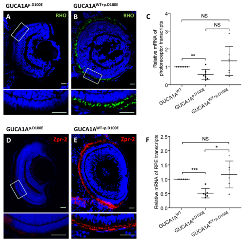
Photoreceptor and RPE defects caused by GUCA1A p.R120L in zebrafish. (A-B, D-E) Both RHO and Zpr-2 staining were diminished in GUCA1Ap.D100E injected zebrafish (A, D), but was re-emerged in the co-injection group (B, E). (C, F) Relative mRNA levels of photoreceptor (C) and RPE (F) characteristic transcripts in GUCA1Ap.D100E and injected groups were decreased when compared with GUCA1AWT injected zebrafish. No statistical significance was noticed between the GUCA1AWT and GUCA1AWT+p.D100E injected zebrafish. |