
- Title
-
FLNC Gene Splice Mutations Cause Dilated Cardiomyopathy.
- Authors
- Begay, R.L., Tharp, C.A., Martin, A., Graw, S.L., Sinagra, G., Miani, D., Sweet, M.E., Slavov, D.B., Stafford, N., Zeller, M.J., Alnefaie, R., Rowland, T.J., Brun, F., Jones, K.L., Gowan, K., Mestroni, L., Garrity, D.M., Taylor, M.R.
- Source
- Full text @ JACC Basic Transl Sci
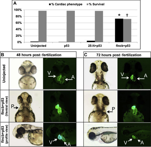
MO Knockdown of flncb in Zebrafish Showing a Cardiac Phenotype (A) Black bars: percentage of surviving embryos with severe cardiac phenotype at 72 h post-fertilization (hpf) for uninjected (n = 411), p53 morpholino (MO)-injected (n = 139), 25-N+p53 control MO-injected (n = 351), and filamin Cb (flncb)+p53 MO-injected (n = 241); *p = 1.04 × 10-83 (for comparison of uninjected heart vs. flncb+p53 MO using chi-square testing). Gray bars: percentage of surviving embryos at 7 days for uninjected (n = 411), p53 MO-injected (n = 139), 25-N+p53 control MO-injected (n = 351), and flncb+p53 MO-injected (n = 164); †p = 1.65 × 10⁻¹⁹ (for comparison of uninjected embryo vs. flncb+p53 MO using chi-square testing). (B) Representative zebrafish heart pictures for embryo uninjected and flncb MO coinjected with p53 MO zebrafish embryos at 48 hpf. The flncb+p53 MO-injected zebrafish show cardiac phenotype (pericardial edema [P]), and GFP hearts showing less heart looping, elongated atrium, pericardial edema, and a truncated ventricle. (C) Representative zebrafish heart pictures for embryo uninjected and flncb MO coinjected with p53 MO zebrafish embryos at 72 hpf. Among the flncb+p53 MO-injected zebrafish, they show cardiac phenotype, and GFP hearts further show less heart looping, elongated atrium, pericardial edema, and a truncated ventricle. |

ZFIN is incorporating published figure images and captions as part of an ongoing project. Figures from some publications have not yet been curated, or are not available for display because of copyright restrictions. PHENOTYPE:
|
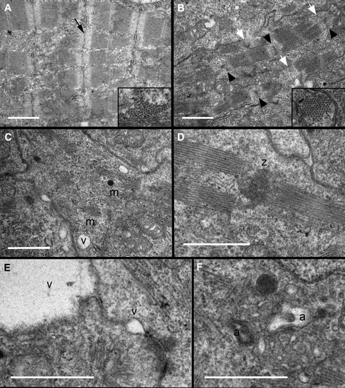
ltrastructural Analysis Indicates Deficiencies in Sarcomere Organization and Z-Disc Formation Transmission electron micrographs of zebrafish hearts at 72 hpf indicate that (A) wild-type embryos had assembled several consecutive sarcomeres with clearly distinct Z-discs (black arrow), whereas flncb+p53 MO hearts (B to F) show evidence of disorganized ultrastructure. (B) Most Z-discs were diffusely stained, irregular in shape (black arrowheads), or seemingly absent (white arrowheads). Insets: Cross section through myofilament bundles indicated a normal primary organization of thick and thin filaments in hexagonal lattices in flncb-depleted hearts. (C) Sarcomere arrangement in myofibrils (m) was often nonconsecutive, with (D) multiple sarcomeres sometimes adjoined to a mass of Z-disc-like material (z). (E) Small vacuoles (v) were often present between or near the plasma membranes of adjacent cardiomyocytes. These vacuoles could be large, creating a gap between adjacent cells. (F) Autophagic vesicles (a) were observed. Scale bar: 1 μmol/l. Abbreviations as in Figure 4. PHENOTYPE:
|
|
ZFIN is incorporating published figure images and captions as part of an ongoing project. Figures from some publications have not yet been curated, or are not available for display because of copyright restrictions. PHENOTYPE:
|