
- Title
-
The seahorse genome and the evolution of its specialized morphology
- Authors
- Lin, Q., Fan, S., Zhang, Y., Xu, M., Zhang, H., Yang, Y., Lee, A.P., Woltering, J.M., Ravi, V., Gunter, H.M., Luo, W., Gao, Z., Lim, Z.W., Qin, G., Schneider, R.F., Wang, X., Xiong, P., Li, G., Wang, K., Min, J., Zhang, C., Qiu, Y., Bai, J., He, W., Bian, C., Zhang, X., Shan, D., Qu, H., Sun, Y., Gao, Q., Huang, L., Shi, Q., Meyer, A., Venkatesh, B.
- Source
- Full text @ Nature
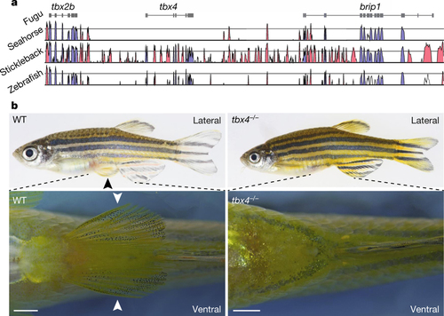
Pelvic fin loss in H. comes is associated with loss of tbx4. a, Vista plot of conserved elements in the tbx2b-tbx4-brip1 syntenic region in fugu (reference genome), seahorse (H. comes), stickleback and zebrafish showing that tbx4 is missing from this locus in seahorse. The blue and red peaks represent conserved exonic and non-coding sequences, respectively. b, Lateral (top) and ventral view (bottom) of wild-type (WT) and a representative (one out of five) F3 homozygous tbx4-null mutant (tbx4−/−) zebrafish. Bottom panel shows a close-up of the pelvic region (dashed lines indicate the approximate zoom region). Scale bar, 1 mm. Pelvic fins are indicated with black or white arrowheads in the wild-type fish. Homozygous tbx4-null mutants entirely lack pelvic fins without showing any other gross morphological defects. PHENOTYPE:
|

ZFIN is incorporating published figure images and captions as part of an ongoing project. Figures from some publications have not yet been curated, or are not available for display because of copyright restrictions. PHENOTYPE:
|
|
ZFIN is incorporating published figure images and captions as part of an ongoing project. Figures from some publications have not yet been curated, or are not available for display because of copyright restrictions. EXPRESSION / LABELING:
|
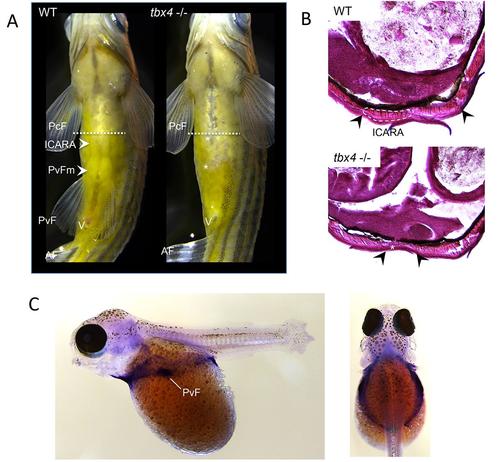
Ventral body wall defects in tbx4 mutant zebrafish. a) ventral view of wildtype and tbx4 mutant fish. Wildtype fish show a distinct pattern of ventral musculature with pronounced bulges at the position of the pelvic girdle (pelvic fin musculature; PvFm) and more anteriorly the musculus infracarinalis anterior(ICARA). In tbx4 mutant fish these muscles are not observed and these fish show a slightly translucent groove along their midline indicative of defects in the formation of their body wall (muscle absence indicated with asterisks). b) Cryosectioning (section shown approximately at the position of the dashed lines indicated in panel aof this figure) shows absence or strong malformation of the musculus infracarinalis anteriorin the mutant leaving a gap in the body wall (musculus infracarinalis anteriorindicated with a dashed outline in the wildtype). Black arrowheads indicate the ventral position of the hypaxial muscles. c) In situhybridization for tbx4in embryos of a cichlid fish (Astatotilapia burtoni) -left panel lateral view, right panel dorsal view. Tbx4 shows expression in a much wider domain than the pelvic fin (indicated PvF) and is also expressed in a ring around the embryo in cells that will contribute to the future ventral midline. This wider expression domain is consistent with the phenotype observed in the tbx4mutant fish. Abbreviations: Pcf; pectoral fin, PvF; pelvic fin, PvFm; pelvic fin musculature, AF; anal fin, ICARA; musculus infracarinalis anterior. PHENOTYPE:
|