
- Title
-
Planar cell polarity genes Frizzled3a, Vangl2, and Scribble are required for spinal commissural axon guidance
- Authors
- Sun, S.D., Purdy, A.M., Walsh, G.S.
- Source
- Full text @ BMC Neurosci.
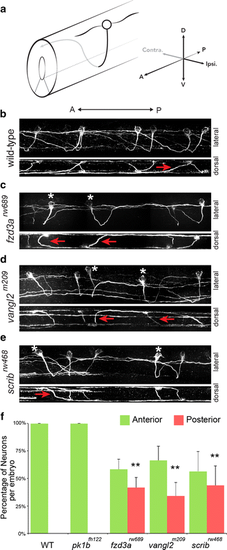
PCP genes are required for anterior pathfinding of CoPA axons after midline crossing. a Illustration of a CoPA neuron in the spinal cord. Black line represents ipsilateral ventral projection, Gray line indicates dorso-anterior trajectory after midline crossing. CoPA neurons have two major dendrites, one projecting anteriorly, and the other projecting posteriorly, b confocal micrographs of 3A10 immunofluorescence shows the pathfinding of CoPA axons in multiple segments of the spinal cord in wild-type (WT) embryos. In the lateral view (upper panel), all CoPA axons project dorso-anteriorly after midline crossing. Anterior is to the left, dorsal is up. In the lower panel, a dorsal view of the same spinal cord shows the midline crossing trajectory of CoPA axons (red arrows), c–e in fzd3 rw689 , vangl2 m209 , and scrib rw468 mutant embryos, approximately half of CoPA axons fail to turn anteriorly after crossing the midline. Asterisks mark affected CoPA cells that turn posteriorly, inappropriately. Lateral views of the same spinal cords show that affected CoPA axons still cross the midline (red arrows), g quantification of anterior–posterior trajectories of CoPA axon after midline crossing per embryo. Error indicates SD. **p < 0.01 versus WT |
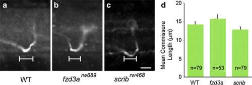
Commissure formation is not affected by loss of fzd3a or scrib. a–c Confocal micrographs showing lateral view of commissures from individual CoPA axons in WT, fzd3a rw689 , and scrib rw468 embryos. Anterior is to the left, dorsal is up, d Measurements of distance traveled by axon within the midline of the spinal cord in the anterior–posterior axis. No significant difference was found in the length of commissures in WT, fzd3a rw689 , and scrib rw468 embryos. n number of neurons counted. A two-tailed Student’s t test was used for statistical analysis. Error indicates SEM |
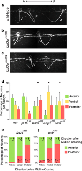
Pre-crossing commissural axon pathfinding is influenced by PCP components. a–c Confocal micrographs of CoPA axon pathfinding in WT and fzd3a rw689 embryos illustrating examples of CoPA axon trajectories. Anterior is to the left, dorsal is up. a CoPA axons have an anterior–posterior bias as they extend ventrally towards the midline. The majority of CoPA axons traverse ventro-anteriorly (A), straight ventrally (V), while a small proportion extend ventro-posteriorly (P). All CoPAs turn anteriorly by the time they have reached the contralateral spinal cord in wild-type embryos, b in fzd3a rw689 mutants, examples of pre-crossing CoPA axons that project ventro-posteriorly (P) and continue posteriorly after midline crossing, and a small number that project ventro-anteriorly before turning posteriorly after midline crossing, c in fzd3a rw689 mutants, pre-crossing CoPA axons that project straight ventrally (V) are more likely to make random A–P guidance as post-crossing fibers, d quantitation of the midline crossing points relative to cell body position among genotypes per embryo (*p < 0.05; ♦p < 0.01; Pearson Chi square test). e, f distribution of post-crossing axon trajectory relative to the pre-crossing directional bias in fzd3a rw689 and scrib rw468 mutants. Superimposed on the graph are representations of the appearance of CoPA trajectories |
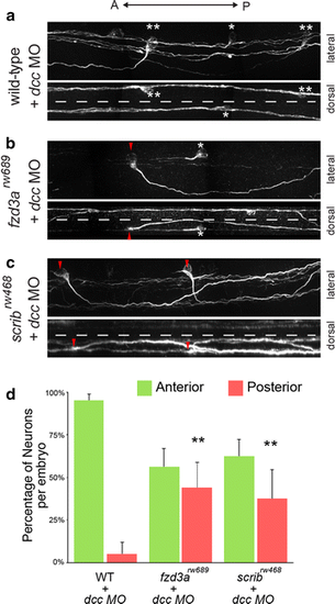
Anterior guidance of CoPA axons in the absence of midline crossing is dependent on PCP proteins. a CoPA axons fail to cross the midline in dcc knockdown embryos yet still undergo anterior growth. In the lateral view, asterisk indicates affected CoPA that pathfinds anteriorly with no ventral growth. Double asterisk indicates affected CoPA that displays weak ventral extension followed by projection dorsally and anteriorly without crossing the midline. Dorsal view of the same spinal cord demonstrates that affected CoPAs are prevented from crossing the midline (dotted line), b, c CoPA axons in fzd3a rw689 and scrib rw468 embryos injected with dcc morpholinos also fail to cross the midline. Red arrowhead indicates uncrossed CoPA axons that grow posteriorly inappropriately in dcc-deficient fzd3a rw689 and scrib rw468 embryos. Asterisk indicates uncrossed CoPA axon that projects anteriorly. Dorsal views of the same spinal cords demonstrate that posteriorly projecting CoPAs fail to cross the midline (dotted line), d quantification of the anterior or posterior trajectory of uncrossed CoPA neurons in dcc-deficient WT, fzd3a rw689 , and scrib rw468 embryos. Error indicates SD. **p < 0.01 versus WT + dcc MO |