FIGURE SUMMARY
Title

The zebrafish as a model for studying neuroblastoma

Authors
Corallo, D., Candiani, S., Ori, M., Aveic, S., Tonini, G.P.
Source
Full text @ Cancer Cell Int.

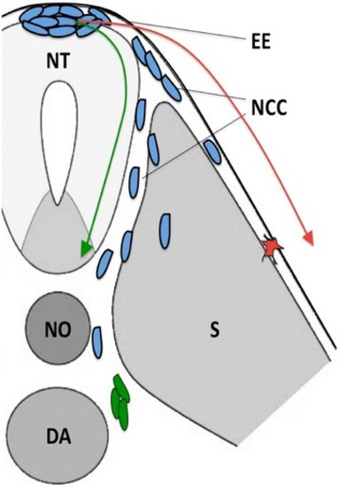
Patterns of NCC migration in zebrafish. Schematic cartoon depicting the two main migratory pathways of NCCs during embryonic development (transverse section of a vertebrate embryo). NCCs (blue) follow the ventromedial pathway (green arrow) and migrate between the neural tube and somites and then bilaterally reach the dorsal portion of the dorsal aorta, where they differentiate into sympathoadrenal progenitors (green). NCCs that migrate dorsolaterally (red arrow) between somites and the overlying ectodermal epithelium give rise to pigment cells (red). NCCs neural crest cells, EE ectodermal epithelium, NT neural tube, NO notochord, DA dorsal aorta, S somite

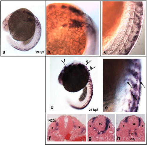
Expression of crestin in neural crest cells (NCCs) during zebrafish development. aCrestin is expressed in premigratory NCCs in wild-type embryos at 19 hpf. b, c Higher magnification images of the embryo in a, showing the streams of migrating NCCs labeled with the crestin probe. d, e At 24 hpf, trunk migratory NCCs labeled by crestin (arrowheads) gradually migrated toward the ventral dorsal aorta. fh Cross sections through the levels (f, g, h) shown in d. Streams of trunk NCCs defined by crestin expression migrate between the somite and the neural tube. da dorsal aorta; n notochord; s somite; sc spinal cord

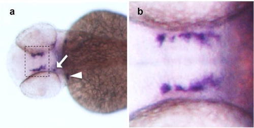
Expression of TH by fully differentiated sympathetic neurons in zebrafish. a Dorsal view of a wild-type embryo probed at 48 hpf by in situ hybridization with TH. At this developmental stage, TH mRNA is expressed by dopaminergic neurons of the ventral diencephalon (hatched box), arch-associated catecholaminergic neurons (arrowhead) and neurons constituting the locus coeruleus (arrow). b Higher magnification of dopaminergic neurons stained with the TH probe

Acknowledgments
This image is the copyrighted work of the attributed author or publisher, and ZFIN has permission only to display this image to its users. Additional permissions should be obtained from the applicable author or publisher of the image. Full text @ Cancer Cell Int.