
- Title
-
Characterization of Zebrafish Models of Marinesco-Sjögren Syndrome
- Authors
- Kawahara, G., Hayashi, Y.K.
- Source
- Full text @ PLoS One

Altered skeletal muscle in sil1-morpholino injected fish. A: Left panels are pictures under bright field, and left panels are birefringence assay (BR). Abnormal structure of muscle is obvious in sil1 morpholino injected fish (MO1 and MO2) compared to control (CMO) under birefringence assay. B: Histogram of the percentage of normal, affected, and dead fish by morpholino-injection. Injection of 3 ng of MO1 or MO2 resulted in approximately 39.0% and 21.8% of injected embryos exhibiting reduced birefringence, respectively. C: Restoration of sil1 morphant with co-injection of fish sil1 mRNA (50 pg). Histogram of the percentage of dead, affected fish of morphants and recovered fish. Co-injection of zebrafish sil1 mRNA with each MO (3 ng) rescued the phenotypes. White bar shows normal %, gray shows affected % and black shows dead fish %. Non: non injected fish, MO1: MO1 injected fish, MO2: MO2 injected fish. PHENOTYPE:
|
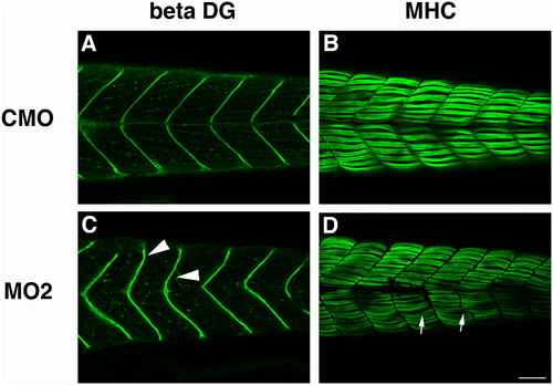
Immunohistochemistry of skeletal muscle tissue of morpholino-injected fish. Immunostaining of CMO injected fish (A, B) and sil1 morphant (MO2) injected fish (C, D) with antibodies against beta-dystroglycan (beta-DG) (A, C) and myosin heavy chain (MHC) (B, D). Beta-dystroglycan expression at the myosepta of MO2 injected 4 dpf embryos is misshapen and has a less clear v-shaped structure. Staining with anti-MHC indicated that formation of myofibers is disturbed in MO2. Arrowheads indicate the disturbance of myosepta in MO2-injected fish. Bar: 100 μm. Arrows indicate the abnormal structure of myofibers. |

Smaller sized eyes in sil1-morpholino injected fish (4 dpf). The diameter of eyes in MO1 or MO2 injected 4 dpf embryos are smaller than those of CMO injected embryos. Co-injection of zebrafish sil1 mRNA with each MOs rescues the eye size. Eye size is measured under a dissection scope with DP controller software and ImageJ. (A) wild type, (B) CMO injected fish, and (C) MO2 injected fish, showing white lines. Measurement of the diameter of eyes (D: MO1, E: MO2). (F): Restoration of eye diameter of sil1 morphant with co-injection of fish sil1 mRNA. Single asterisk indicates Student’s t-test p = 1.25E-07 (MO1; n = 19, CMO; n = 14), double asterisks indicate p = 7.36E-08 (MO2; n = 16, CMO; n = 28) and triple asterisks indicate p = 0.00441 (MO2; n = 19, MO2+sil1 mRNA 50 pg; n = 14, CMO; n = 14). PHENOTYPE:
|

Staining of purkinje cells with anti-purvalbumin in cerebellar area. The number of purkinje cells detected with anti-parvalbumin shows reduced number of positive cells in MO1 or MO2 injected embryos. (A, B and C): CMO injected fish, (D, E and F): MO1 injected fish, (G, H and I): MO2 injected fish. (A, D and G: Staining Purkinje cell. (B, E and H): DAPI, nuclei. (C, F and I): Merged images. (J): Increased number of Purkinje cells in sil1 morphants with co-injection of fish sil1 mRNA (50 pg) from 23.1% (MO2, n = 13) to 64.3% (MO2+sil1 mRNA, n = 14). PHENOTYPE:
|

ZFIN is incorporating published figure images and captions as part of an ongoing project. Figures from some publications have not yet been curated, or are not available for display because of copyright restrictions. PHENOTYPE:
|