
- Title
-
PHACTR1 Is a Genetic Susceptibility Locus for Fibromuscular Dysplasia Supporting Its Complex Genetic Pattern of Inheritance
- Authors
- Kiando, S.R., Tucker, N.R., Castro-Vega, L.J., Katz, A., D'Escamard, V., Tréard, C., Fraher, D., Albuisson, J., Kadian-Dodov, D., Ye, Z., Austin, E., Yang, M.L., Hunker, K., Barlassina, C., Cusi, D., Galan, P., Empana, J.P., Jouven, X., Gimenez-Roqueplo, A.P., Bruneval, P., Hyun Kim, E.S., Olin, J.W., Gornik, H.L., Azizi, M., Plouin, P.F., Ellinor, P.T., Kullo, I.J., Milan, D.J., Ganesh, S.K., Boutouyrie, P., Kovacic, J.C., Jeunemaitre, X., Bouatia-Naji, N.
- Source
- Full text @ PLoS Genet.

phactr1 modulation in zebrafish affects vascular dimensions and patterning. Two-dimensional projections obtained from z-series confocal images in the trunk of control and phactr1 knockdown (KD) zebrafish embryos (two representative images per condition). Green represents the vascular endothelium as marked by EGFP. Greyscale represents the corresponding DIC bright field image of the fish trunk. DA: Dorsal aorta, SV: Segmental vessel, PCV: Posterior cardinal vein. Quantification of inner vascular diameter for the dorsal aorta (DA), posterior cardinal vein (PCV) and caudal artery (CA). (*) represents P<0.05. PHENOTYPE:
|
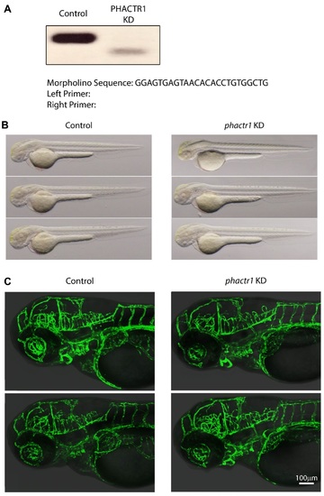
Phenotypic evaluation of phactr1 knockdown zebrafish. (A) RT-PCR evaluation of splice alteration observed following microinjection of phactr1 morpholino (PHACTR1 KD). (B) Brightfield micrographs of overt morphology at 60 hours post fertilization. (C) Two-dimensional projections obtained from z-series confocal images in the head and trunk of control and phactr1 knockdown zebrafish embryos. Green represents the vascular endothelium as marked by EGFP. Greyscale represents the corresponding DIC brightfield image of the fish head and trunk region. HPV indicates the relative position of the developing hepatic portal vein PHENOTYPE:
|