
- Title
-
eduSPIM: Light Sheet Microscopy in the Museum
- Authors
- Jahr, W., Schmid, B., Weber, M., Huisken, J.
- Source
- Full text @ PLoS One
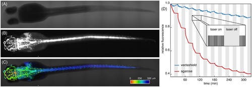
72 hpf zebrafish embryos expressing GFP in the vascular system. Prior to imaging, all zebrafish embryos were fixed and their fluorescence signal was recovered using booster-GFP. (A): Transmission image and (B): fluorescence image of vasculature of the same embryo. (C): Overlay of fluorescence and transmission data. Fluorescence data was 3D-rendered and colour coded for depth. Dataset of 75planes, colour bar, 500 m long. (D): Fluorescence signal of the sample embedded in vectashield (blue) and agarose (red). The sample was illuminated every 30s for 15min, followed by a 15min dark period (inset). Photobleaching dominated during bright periods. For mounting in vectashield, overall photobleaching was greatly reduced and even negligible during dark periods. |