
- Title
-
The role of Rho kinase (Rock) in re-epithelialization of adult zebrafish skin wounds
- Authors
- Richardson, R., Hammerschmidt, M.
- Source
- Full text @ Small GTPases
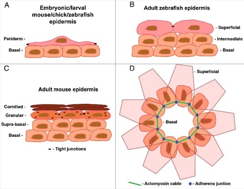
(A-C) Representations of cross sections through the epidermis at different time-points and in the different models used for wound healing studies. (A) In embryonic mice and chicks the epidermis is bilayered at the stages used for embryonic wound closure experiments (E12 in mouse, E6 in chick). At these stages the epidermis consists of an inner basal layer and an outer covering of flattened periderm cells. The epidermis of late embryonic and larval zebrafish is very similar in structure and function. (B) In adult zebrafish the epidermis becomes stratified consisting of 2 to 3 layers of basal and intermediate cells and an outer superficial layer. This resembles the epidermis of mid-gestation mice (E13-E15) and chicks (E15-E17) of corresponding developmental stages. (C) The epidermis of adult mice and chicks is also stratified consisting of 4 distinct layers: the innermost basal layer, the suprabasal layer, the granulation layer where terminal differentiation commences and the outermost cornified layer consisting of terminally differentiated cornified envelopes. (D) Representation of a dorsal view of the bilayered epidermis of an embryonic chick or mouse depicting the actomyosin cable form of wound closure. For simplicity only the LE cells of the basal epidermis are shown. The first row of superficial periderm cells are depicted as semi-translucent to allow vizualization of the basal cells beneath. |
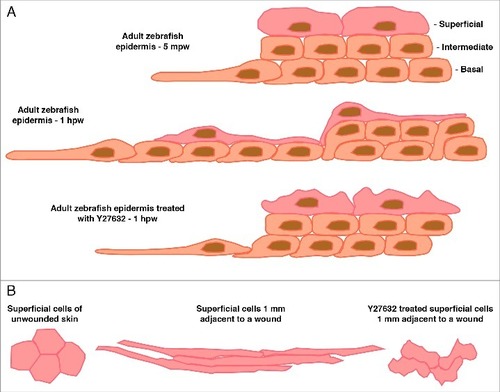
(A) Representation of a cross section through the epidermis at the wound edge in an adult zebrafish 5 minutes post-wounding (mpw), one hour post-wounding (hpw), or 1 hpw following Rock inhibition. The epidermis initially consists of 2-3 layers of inner basal and intermediary cells and a layer of superficial cells. Within 5 mpw, the LE basal cells have begun to make lamellipodial protrusions and elongate onto the denuded area. By 1 hpw directed radial intercalations of intermediary cells into the basal layer have occurred producing a single layer of inner cells; the superficial cells have elongated to ensure coverage of the increased basal layer area; the LE cells and several cells behind are still protrusive and have undergone some cell elongation, collectively allowing the whole epidermal sheet to move forward to cover the wound. Following Rock inhibition the processes of radial intercalation and coordinated elongation are severely affected resulting in reduced forward movement even though LE protrusions are maintained. (B) Dorsal view outlines of 4 individual superficial cells taken from images from adult zebrafish at the indicated distance from a full-thickness wound (wound is to the left). In unwounded epidermis, the superficial cells are hexagonal in shape. Following wounding the superficial cells elongate in the direction of the wound to up to 10-times their original diameter. Following Rock inhibition the superficial cells change shape but in an uncoordinated manner resulting in many random shapes. |