
- Title
-
Altered RNA metabolism due to a homozygous RBM7 mutation in a patient with spinal motor neuropathy
- Authors
- Giunta, M., Edvardson, S., Xu, Y., Schuelke, M., Gomez-Duran, A., Boczonadi, V., Elpeleg, O., Müller, J.S., Horvath, R.
- Source
- Full text @ Hum. Mol. Genet.
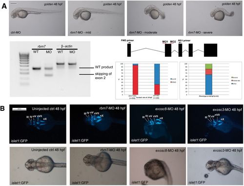
Morphological defects caused by injection of rbm7 MO (A). Mild phenotype fish show slight developmental delay, and shorter body length. Rostrally the head is smaller than control fish while caudally the head looks slightly swollen. Body shape in the moderate phenotype looks compromised and head oedema increased, sporadically we could observe heart oedema as well. Body shape of the severe fish is completely disrupted, swelling of the head is more severe, and we detected smaller forehead and eyes compared with control fish (Scale bar = 200 μm). On the bottom left: RT-PCR of zebrafish cDNA shows efficacy of morpholino against In1-Ex2 boundary. Position of morpholinos is explained in the graph on the right. Below: graphs showing mortality and phenotype rates at 24 hpf. (B) Hindbrain defects in morphant fish. Cranial motor neurons nuclei are well defined in 5 nuclei in wild type fish: Oculomotor neurons (III), Trochlear neurons (IV), Trigeminal neurons (V), Facial neurons (VII), Vagal neurons (X). rbm7-MO seems to have a milder effect on cranial motor-neurons development and to affect predominantly vagal nucleus (nX) which result shorter than controls. Hindbrain structures in exosc8-MO are more generally affected the moderate phenotype fish. Only nX is partially preserved. Exosc3-MO affects predominantly nVII (facial neurons) (Scale bar = 200 μm). PHENOTYPE:
|
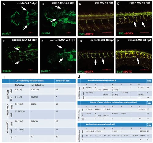
Confocal microscopy analysis of PCs (A,B,E,F) and neuromuscular junctions (C,D,G,H) in zebrafish upon injection of three different MOs. PCs fail to differentiate in rbm7-MO (B), exosc8-MO (E) and exosc3-MO (F) zebrafish compared with ctrl-MO fish (A). The percentage of fish with cerebellar defects varies significantly between morphants, reflecting what observed in patients. Motor axon growth is defective in all three morphants (D,G,H); arrows point at abnormally short motor axons. Interestingly, only in exosc8-MO the motor neuron axon branches in close proximity of the spinal cord instead of more ventrally as in the ctrl-MO fish (G). Scale bar = 100 μm. We show the quantity and respective percentage of fish with cerebellar defects (I). Axonal defects in different morphant and phenotypical classes (only mild and moderate phenotypes were considered for this analysis) (J). The number of defective structures increases with the severity of the phenotype. |

ZFIN is incorporating published figure images and captions as part of an ongoing project. Figures from some publications have not yet been curated, or are not available for display because of copyright restrictions. EXPRESSION / LABELING:
PHENOTYPE:
|
|
ZFIN is incorporating published figure images and captions as part of an ongoing project. Figures from some publications have not yet been curated, or are not available for display because of copyright restrictions. PHENOTYPE:
|