
- Title
-
Sodium Selenite Acts as an Otoprotectant against Neomycin-Induced Hair Cell Damage in a Zebrafish Model
- Authors
- Chang, J., Choi, J., Rah, Y.C., Yoo, M.H., Oh, K.H., Im, G.J., Lee, S.H., Kwon, S.Y., Park, H.C., Chae, S.W., Jung, H.H.
- Source
- Full text @ PLoS One
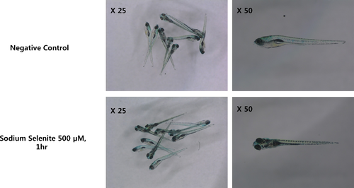
The effect 1hr exposure of 500 μM sodium selenite on zebrafish development. The viability of 6 days post-fertilization zebrafish, three days after the treatment with 500 μM sodium selenite for 1 hr, was evaluated. There were no noticeable developmental differences between zebrafish treated with sodium selenite compare to those of the negative control group. PHENOTYPE:
|
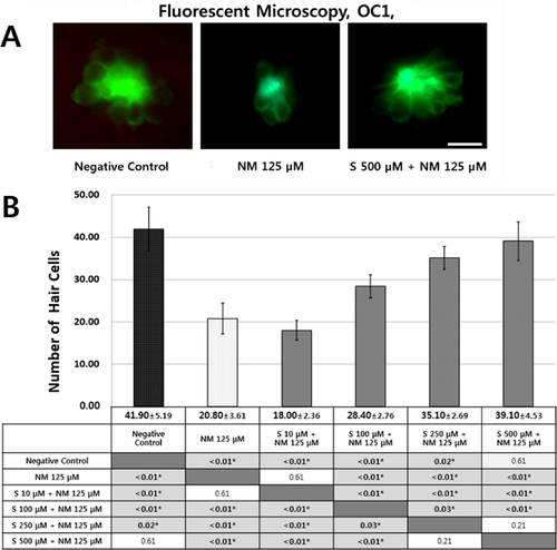
Fluorescent microscopy (OC1, ×40) and quantitative assay for hair cell damage in the five and six days post-fertilization (dpf) transgenic zebrafish (Brn3C: EGFP). The 5 and 6-dpf transgenic zebrafish were treated simultaneously with 125 μM neomycin and various concentrations (10, 100, 250, and 500 μM) of sodium selenite for 1h. Treatment with neomycin resulted in a significant decrease in the number of hair cells, but 500μM sodium selenite most attenuated the hair cell damage (A). Hair cells from four neuromasts (SO1, SO2, O1, and OC1) were analyzed. The total hair cells of negative control group had 41.90 ± 5.19 cells. Treatment of the transgenic zebrafish with 125 μM neomycin for 1 h significantly decreased the number of hair cells in the neuromast (20.80 ± 3.61 cells). Sodium selenite protected against neomycin-induced hair cell loss in a dose dependent manner. The concentration of 500 μM of sodium selenite had significantly protective effect against neomycin (*: statistically significant) (n = 10 fish per treatment, B). NM 125 μM: 20.80 ± 3.61 cells, p<0.01, compared to negative control; S 10 μM + NM 125 μM: 18.00 ± 2.36 cells, p<0.01 and 0.61, compared to negative control and NM 125 μM, respectively; S 100 μM + NM 125 μM: 28.40 ± 2.76 cells, p<0.01, p<0.01, and p<0.01, compared to negative control, NM 125 μM, and S 10 μM + NM 125 μM, respectively; S 250 μM + NM 125 μM: 35.10 ± 2.69 cells, p = 0.02, p<0.01, p<0.01, and 0.03, compared to negative control, NM 125 μM, S 10 μM + NM 125 μM, and S 100 μM + NM 125 μM, respectively; S 500 μM + NM 125 μM: 39.10 ± 4.53 cells, p = 0.61, p<0.01, p<0.01 p<0.01, and p = 0.21, compared to negative control, NM 125 μM, S 10 μM + NM 125 μM, S 100 μM + NM 125 μM, S 250 μM + NM 125 μM, respectively (B). * statistically significant: Scale bar = 10 μm. NM, neomycin; S, sodium selenite. |
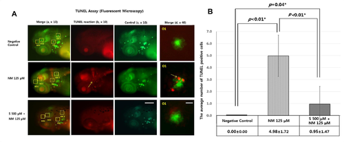
Quantitative analysis of apoptotic cell death by TUNEL assay. TUNEL staining as a quantitative assay was performed to determine the protective effect of sodium selenite against neomycin-induced apoptosis. Apoptotic cells are marked as light red dots in red-colored fish after TUNEL staining as observed under a fluorescent microscope (arrow indicates TUNEL-positive cells in TUNEL reaction (b) column and merge (d) column). Light red dots were not visible in the negative control zebrafish (A). A comparison of the color intensity between the group treated with 125 μM neomycin and the group treated with 500 μM sodium selenite for 1 h showed that the number of TUNEL-positive cells significantly decreased in the 5 and 6-dpf transgenic zebrafish treated with sodium selenite, and that apoptotic cell death of the neuromasts hair cells was attenuated by sodium selenite (green and red-colored fish in the left column, combination of control and TUNEL reaction (a), × 10; red-colored fish in the left middle column, TUNEL reaction (b), × 10; green-colored fish in the right middle column, control for TUNEL reaction (c), × 10; green and red-colored neuromast in the right column, merge for control and TUNEL reaction by high magnification (d), × 40) (A). The concentration of 500 μM of sodium selenite reduced significantly neomycin-induced TUNEL positive cells in four neuromasts (SO1, SO2, O1, and OC1) (*: statistically significant) (B). Negative control: TUNEL positive cells = 0.00 ± 0.00; NM 125 μM: the average TUNEL positive cells, 4.98 ± 1.72; S 500 μM + NM 125 μM: the average TUNEL positive cells = 0.95 ± 1.47; n = 10 fish per treatment, *: statistically significant. (B). Scale bars = 200 μm (c); 20 μm (d). NM, neomycin; S, sodium selenite. |
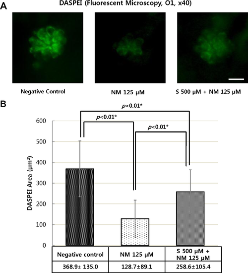
Analysis of hair cell damage by DASPEI assay (×40). The wild type zebrafish were treated with 125 μM neomycin and 500 μM sodium selenite for 1 h. Staining hair cells with DASPEI showed that treatment with 125 μM neomycin caused reduced numbers of hair cells in neuromast. However, the co-treatment with 500 μM sodium selenite protected reducing number of hair cell staining with DASPEI (A). The concentration of 500 μM of sodium selenite preserved significantly the average DASPEI area in four neuromasts (SO1, SO2, O1, and OC1) (n = 20 fish per treatment; *: statistically significant) (B). Negative control: the average DASPEI area = 368.9 ± 135.0 μm2; NM 125 μM: the average DASPEI area = 128.7 ± 89.1 μm2; S 500 μM + NM 125 μM: the average DASPEI area = 258.6 ± 105.4 μm2(B). NM, neomycin; S, sodium selenite. Scale bar = 10 μm. |
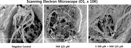
Scanning electron microscopy (SEM, O1, x 10K). The kinocilium (K) and the stereocilia bundles (asterisk) of hair cells in neuromast were clearly visible in the negative control (A). However, when the 5-dpf transgenic zebrafish were treated with 125μM neomycin for 1 h, the kinocilium and the stereocilia bundles were affected and severely disrupted (B). Sodium selenite provided nearly complete protection against neomycin-induced the damage of the kinocilium (K) and the stereocilia bundles (asterisk) in the neuromasts (C). Images were obtained in three 5-dpf zebrafish for each group. Scale bar (at the bottom of each figure, one space) = 3 μm. NM, neomycin; S, sodium selenite. PHENOTYPE:
|
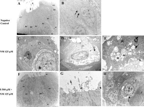
Transmission electron microscopy (TEM). TEM of Normal control (A and B); TEM of zebrafish treated with 125 μM neomycin only (C-E); TEM of zebrafish treated with 125 μM neomycin and 500 μM sodium selenite (F-H). The sterocilia (black asterisk) and the kinocilium (K) from each hair cell are clearly visible (× 8K) (A). A normal-sized mitochondria (arrow, × 25K) (B). The hair cells were severely damaged and showed a condensed nuclei (black asterisk) and pyknotic nuclei (white asterisk) (× 20K) (C). The collapse of the apical surface of neuromasts was typically evident. A hair cell showed extrusion of cytoplasm (black asterisk) and the swollen mitochondria (arrow) (× 20K) (D).Severely damaged hair cells show a severely degenerating cytoplasm (black asterisk), a fragmenting condensed nucleus (white asterisk), and multiple swollen mitochondria (M) (× 30K) (E).When 500 μM sodium selenite was applied, structure of neruomasts were nearly complete protected. Nuclear damage such as condensed cytoplasm and pyknotic nuclei were not showed. (× 5 K) (F). The structure of the stereocilia (black asterisk) and the kinocilium(K) are preserved (× 20 K) (G). The normal-sized nucleus (black asterisk) and mitochondria (arrow) is shown (× 20 K) (H). NM, neomycin; S, sodium selenite; HC, hair cell; SC, supporting cell; CC, crescent cell; MC, mantle cell; BM, basement membrane. Scale bars (at the top or the bottom of each figure, one space) = 5μm (A); 2μm (B); 2μm(C); 1μm (D); 1 μm (E); 2μm (F); 2μm (G); 2μm (H). NM, neomycin; S, sodium selenite. Images were obtained in three 5-dpf zebrafish for each group. PHENOTYPE:
|