
- Title
-
klf2ash317 Mutant Zebrafish Do Not Recapitulate Morpholino-Induced Vascular and Haematopoietic Phenotypes
- Authors
- Novodvorsky, P., Watson, O., Gray, C., Wilkinson, R.N., Reeve, S., Smythe, C., Beniston, R., Plant, K., Maguire, R., M K Rothman, A., Elworthy, S., van Eeden, F.J., Chico, T.J.
- Source
- Full text @ PLoS One
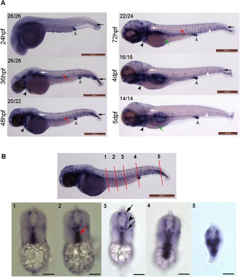
klf2a expression patterns in developing zebrafish embryos. (A) klf2a expression patterns were examined using whole-mount in situ hybridisation. Grey arrowheads indicate cloaca, black arrows indicate cells lateral to the most posterior notochord, white arrows indicate pectoral fin, red arrows indicate trunk vasculature, black arrowheads indicate the cardiac outflow tract, white arrowheads indicate neuromasts and green arrows indicate subintestinal veins (3dpf) or hepatic portal vein (5dpf). Numbers in the top left corners indicate number of embryos with similar staining patterns out of total number of embryos examined. Scale bar = 500μm. (B) Cross sections of a 48hpf wildtype embryo showing klf2a expression in dorsal aorta (red arrow), parachordal vessel (black arrow), intersegmental vessel (ISV) (black arrowhead) and dorsal longitudinal anastomotic vessel (DLAV) (dotted black arrow). Anatomical positions of sections are indicated by the red lines and numbers on the top panel figure with corresponding cross sections images in the bottom panel. Scale bar = 500μm (top panel) and 100μm (bottom panel). |

Comparison of the morphology of wildtype and klf2ash317 mutant embryos. There are no obvious morphological differences between wildtype and klf2ash317 mutant embryos up to 5dpf and also beyond up to adulthood (not shown). Scale bar = 500μm. |

ZFIN is incorporating published figure images and captions as part of an ongoing project. Figures from some publications have not yet been curated, or are not available for display because of copyright restrictions. PHENOTYPE:
|

The vascular anatomy of homozygous klf2ash317 mutants. (A) Formation of the 5th accessory aortic arch (AA5x) connecting the 5th and 6th aortic arch is not affected in homozygous klf2ash317 mutants. Top panel figure shows vascular anatomy of a zebrafish embryo at 3dpf. White rectangle indicates the location of aortic arches. DA denotes dorsal aorta. Bottom panel figures represent dorsal views on aortic arches. White arrows indicate lateral dorsal aortae. White arrowheads indicate AA5x vessels. Scale bar = 200μm. (B) Quantification of endothelial cell nuclei in 4 ISVs closest to the cloaca and corresponding DLAV in WT and klf2ash317 mutants at 3dpf. Scale bar = 70μm. |

Vascular development in vhlhu2117 mutants is unaffected by the klf2ash317 mutation. Wildtype embryos exhibit angiogenesis typical for this developmental stage– 3dpf (left figure, white arrow). Homozygous vhlhu2117 embryos show enlargement of vessels (ISVs and DLAV) with increased tortuosity and looping of the DLAV (middle figure, right arrow). The same vascular phenotype was be observed in vhlhu2117 embryos in a homozygous klf2ash317 background (right figure, red arrow). Confocal images of Tg(fli1:eGFP) zebrafish embryos at 3dpf.. Scale bar = 70μm. PHENOTYPE:
|

Expression of HSC markers runx1 and cmyb is unaffected in homozygous klf2ash317 mutants at 36hpf. Expression patterns of HSC markers runx1 (top panel) and cmyb (bottom panel) do not differ between the wildtype and klf2ash317 mutants at 36hpf. Black arrows indicate the aorta-gonad-mesonephros (AGM) region and red arrows indicate caudal haematopoietic tissue (CHT). Numbers in the top left corners indicate number of embryos with similar staining patterns out of total number of embryos examined. Scale bar = 500μm. EXPRESSION / LABELING:
|

klf2b expression patterns do not differ between wildtype and klf2ash317 mutants. klf2b mRNA is detectable in the developing pectoral fin bud (black arrows). Most of the embryos examined exhibit klf2b mRNA presence on the surface of the embryos representing epidermal cells as described before [17, 48] (red arrowheads). A small proportion of both wildtype and klf2ash317 mutant embryos show vascular staining in the ISVs (red arrows) and/or in the subintestinal veins (green arrows). There were no differences in staining patterns and especially in the level of vascular staining observed between wildtype and klf2ash317 mutant embryos. Figures in bottom left corner of each image indicate the number of embryos with similar staining patterns out of total number of embryos examined. Scale bar = 500μm. EXPRESSION / LABELING:
|

klf4a and biklf/klf4b/klf17 expression patterns do not differ between wildtype and klf2ash317 mutants at 48hpf. klf4a expression was detected in epidermis (black arrows) and pectoral fins (red arrowheads) and biklf/klf4b/klf17 expression was detected in neuromasts of lateral line organ (red arrows) and in hatching glands (black arrowheads) of wildtype embryos and klf2ash317 mutants in keeping with previously published data [17,47] No vascular expression of klf4a or biklf/klf4b/klf17 could be detected in any of the wildtype embryos or klf2ash317 mutants examined. Figures in bottom left corner of each image indicate the number of embryos with similar staining patterns out of total number of embryos examined. Scale bar = 500μm. EXPRESSION / LABELING:
|
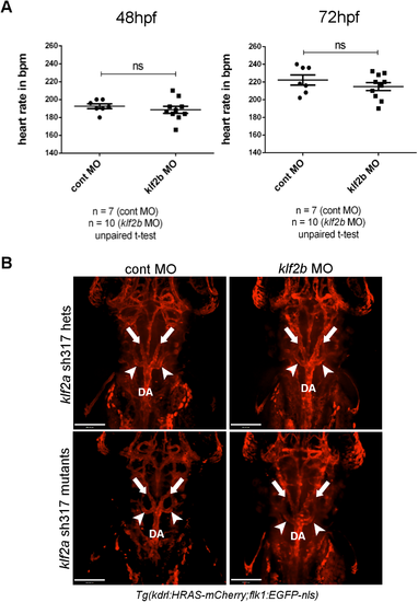
klf2b knockdown does not alter heart rate or AA5x formation in klf2ash317 mutants. (A) klf2b morpholino injection did not affect the heart rate of klf2ash317 mutants at either 48 or 72hpf. (B) AA5x vessel formation (white arrowheads) is intact in klf2b morphants in either a homozygous or heterozygous klf2ash317 mutant background. White arrows indicate lateral dorsal aortae. DA indicates dorsal aorta. Scale bar = 200μm. PHENOTYPE:
|
|
ZFIN is incorporating published figure images and captions as part of an ongoing project. Figures from some publications have not yet been curated, or are not available for display because of copyright restrictions. EXPRESSION / LABELING:
|
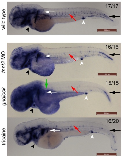
klf2a vascular expression is blood flow-dependent. klf2a is expressed in zebrafish embryonic vasculature of a WT embryo at 48hpf. Cessation of blood flow in the trunk vasculature by an occlusion of proximal aorta in the gridlock mutants (indicated by a green arrow) results in a complete loss of klf2a vascular expression distally to the occlusion. Blockage of embryonic heart contractions by tnnt2 MO results in significantly decreased klf2a vascular expression. Pharmacological inhibition of heart contractions by tricaine from 32 to 48hpf results in significantly decreased klf2a vascular expression. Interestingly tricaine also reduces klf2a expression in the heart region and in the cells lateral to the most posterior notochord. Red arrows indicate trunk vasculature, black arrows indicate the cells lateral to most posterior notochord, white arrows indicate pectoral fins, black arrowheads indicate the cardiac outflow tract and white arrowheads indicate cloaca. Numbers in top right corners indicate the number of embryos with similar staining pattern out of all embryos examined. klf2a riboprobe used. Scale bar = 500μm. |
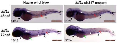
klf2a expression patterns in klf2ash317 mutants. No differences in klf2a WISH staining patterns were detected in klf2ash317 mutant embryos when compared to wildtype counterparts at examined time points indicating the absence of nonsense-mediated mRNA decay (NMD) in klf2ash317 mutants. Grey arrowheads indicate cloaca, black arrows indicate cells lateral to the most posterior notochord, white arrows indicate pectoral fin, red arrows indicate trunk vasculature, black arrowheads indicate the cardiac outflow tract, white arrowheads indicate neuromasts and green arrows indicate subintestinal veins. Numbers in the bottom left corners indicate number of embryos with similar staining patterns out of total number of embryos examined. Scale bar = 500μm. |
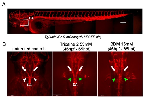
Formation of the AA5x vessel in blood flow dependent. (A) Vascular anatomy of a zebrafish embryo at 3dpf. White rectangle indicates the location of aortic arches. DA denotes dorsal aorta. Scale bar = 200μm. (B) Formation of AA5x vessels in unaffected in the WT embryos (untreated controls) as indicated by the white arrowheads. White arrows point at lateral dorsal aortae. AA5x vessel is missing in embryos treated with tricaine (middle panel) and with BDM (right figure) which both prevent blood flow (green arrowheads). Formation of lateral dorsal aortae is unaffected (white arrows). These findings confirm the previously published data [18]. Scale bar = 70μm. PHENOTYPE:
|
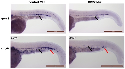
Expression of HSC markers runx1 and cmyb is blood flow dependent. Expression patterns of HSC markers runx1 (top panel) and cmyb (bottom panel) is significantly diminished in the tnnt2 morphants which lack blood flow confirming previously published data [19]. Black arrows indicate the aorta-gonad-mesonephros (AGM) region and red arrows indicate caudal haematopoietic tissue (CHT). Numbers in the top left corners indicate number of embryos with similar staining patterns out of total number of embryos examined. Scale bar = 500μm. |