
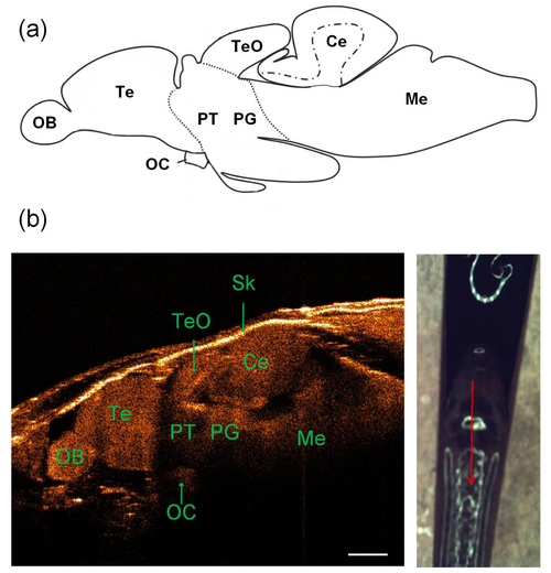
(a) Schematic of the lateral view of adult zebrafish brain (the top row) [27]. (b) A representative sagittal SD-OCT image of the adult zebrafish brain (bottom left) along the red profile as shown in the photograph (bottom right): OB, olfactory bulb; OC, optic commissure; Te, telencephalon; TeO, tectum opticum; Ce, cerebellum; Me, medulla; PG, preglomerular complex; PT, posterior tuberculum; Sk, skull. The scale bar is 500 µm. |
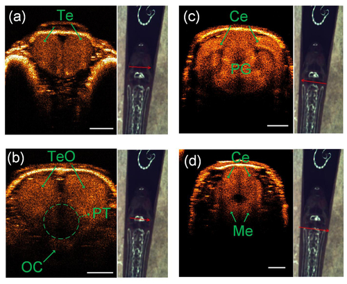
Four different coronal SD-OCT images [(a) to (d)] at four different labeled positions on the head of the same adult zebrafish (see arrow locations and direction): OC, optic commissure; Te, telencephalon; TeO, tectum opticum; Ce, cerebellum; Me, medulla; PG, preglomerular complex; PT, posterior tuberculum. The scale bar is 500 µm. |
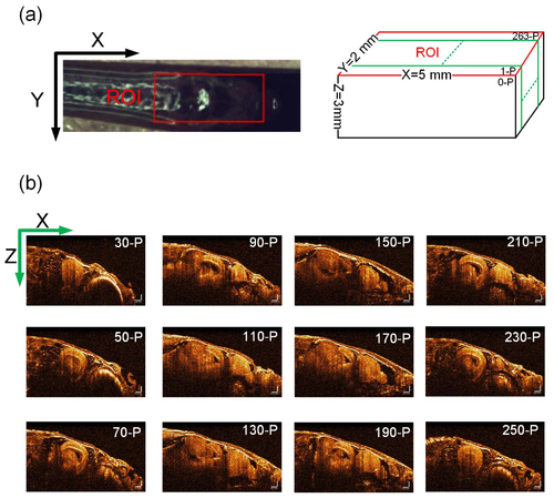
(a) Region of interest (ROI) was labeled with a red rectangle in the photograph that could cover the whole brain. (b) Sequence of sagittal SD-OCT images of the adult zebrafish brain. The scale bar is 250 µm and is same for all the images. |
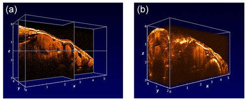
3D view of the reconstructed adult zebrafish brain in the cross-sectional form (a, Visualization 1) and volumetric form (b, Visualization 2). |
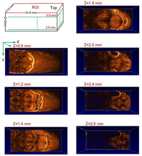
Characterization of the adult zebrafish brain from the horizontal view based on the reconstructed 3D SD-OCT image of the same region of interest (ROI) as shown in Fig. 4 (a) with different imaging depths along Z axis with z = 0.8 mm, 1.2 mm, 1.4 mm, 1.8 mm, 2.0 mm, 2.4 mm and 2.6 mm, respectively. |
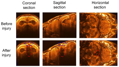
Characterization of the adult zebrafish brain before (the first row) and after (the second row) brain injury. Columns one to three separately showed the coronal, sagittal and horizontal section of the 3D SD-OCT images. |