
- Title
-
Myocardial NF-κB activation is essential for zebrafish heart regeneration
- Authors
- Karra, R., Knecht, A.K., Kikuchi, K., Poss, K.D.
- Source
- Full text @ Proc. Natl. Acad. Sci. USA
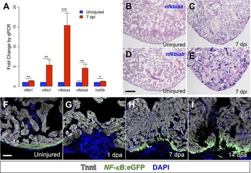
NF-KB activity is induced by injury and during regeneration. (A) qPCR for NF-KB target genes in regenerating ventricles at 7 dpi in cmlc2:CreER; βact2:RS-DTA animals treated with tamoxifen (n = 6) compared with ventricles from cmlc2:CreER; βact2:RS-DTA animals treated with vehicle (n = 6). Error bars indicate SE. *P < 0.05, **P < 0.01, ***P < 0.001, Student’s t test, two-tailed. Each replicate consists of a pool of 2–3 ventricles. (B–E) In situ hybridization for nfkbiaa or nfkbiab on sections from ventricles of cmlc2:CreER; βact2:RS-DTA animals treated with vehicle (B and D) or tamoxifen (C and E). Violet staining indicates expression. (F–I) Time course of NF-KB:eGFP induction during regeneration in sections from an uninjured ventricle, a 1 dpa ventricle, a 7 dpa ventricle, and a 14 dpa ventricle. Ventricles are counterstained with an antibody against Tnnt (gray) to delineate cardiomyocytes. (Scale bars: 100 µm.) |
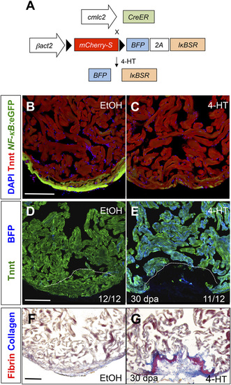
NF-KB is required for heart regeneration. (A) Schematic of βact2:RS-IKBSR transgene. (B and C) Section images of cmlc2:CreER; βact2:RS-IκBSR; NF-KB:eGFP hearts 60 d after treatment (dpt) with vehicle (n = 12) or 4-HT (n = 12). (D and E) Section images of cmlc2:CreER; βact2:RS-IKBSR ventricles at 30 d after amputation after treatment with vehicle or 4-HT. Images are stained for troponin to denote cardiac muscle. Dashed line indicates approximate resection plane. (F and G). Section images of cmlc2:CreER; βact2:RS-IKBSR ventricles at 30 d after amputation after treatment with vehicle or 4-HT. Images are stained with acid fuchsin orange G (AFOG) to identify scar (blue) and fibrin (red). (Scale bars: 100 µm.) |
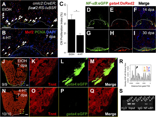
NF-KB modulates cardiomyocyte proliferation and dedifferentiation. (A and B) Section images from cmlc2:CreER; βact2:RS-IκBSR hearts treated with vehicle or 4-HT at 7 dpa. Sections are stained for Mef2 (red) and PCNA (green). Arrowheads indicate Mef2+/PCNA+ nuclei. Dashed line indicates approximate resection plane. (C) Quantification of cardiomyocyte proliferation in cmlc2:CreER; βact2:RS-IκBSR hearts treated with vehicle (n = 15) or 4-HT (n = 17) at 7 dpa. Error bars indicate SEM. *P = 0.004, Mann–Whitney test. (D–I) Sections from NF-KB:eGFP; gata4:DsRed2 hearts at 14 and 30 dpa. (J–Q) Section images from cmlc2:CreER; βact2:RS-IκBSR; gata4:eGFP hearts treated with vehicle (n = 9) or 4-HT (n = 10) at 7 dpa. Inset shows unique high-magnification images for Tnnt, eGFP, and a merge of both channels. (R) Plot for density of putative NFκB1 sites in the gata4 promoter. seqLogo is for the consensus NFκB1 binding site used for analysis. Arrow indicates the region of the promoter with highest density of NFκB1 sites. (S) ChIP-PCR for the region of the gata4 promoter highlighted by arrowhead in R after immunoprecipitation for NFKB1 from cardiac chromatin extracts of cmlc2:CreER; βact2:RS-IκBSR fish treated with vehicle or 4-HT at 7 dpa. (Scale bars: 100 µm.) |
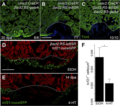
NF-κB has pleiotropic inhibitory effects on heart regeneration. (A–C) Section images of cmlc2:CreER; βact2:BS-gata4, cmlc2:CreER; βact2:RS-IκBSR, and cmlc2:CreER; βact2:BS-gata4; βact2:RS-IκBSR ventricles at 30 dpa after treatment with 4-HT. Images are stained for Tnnt to demarcate cardiac muscle. Dashed line indicates approximate resection plane. (D and E) Tilescan images of cmlc2:CreER; βact2:RS-IκBSR; tcf21:nuceGFP ventricles at 14 dpa after treatment with vehicle or 4-HT. Images are stained for Tnnt to denote cardiac muscle. (F) Quantification of tcf21+ cell responses in cmlc2:CreER; βact2:RS-IκBSR hearts treated with vehicle (n = 10) or 4-HT (n = 12) at 14 dpa. Error bars indicate SE. *P < 0.05, Student’s t test, two-tailed. (Scale bars: 100 µm.) |
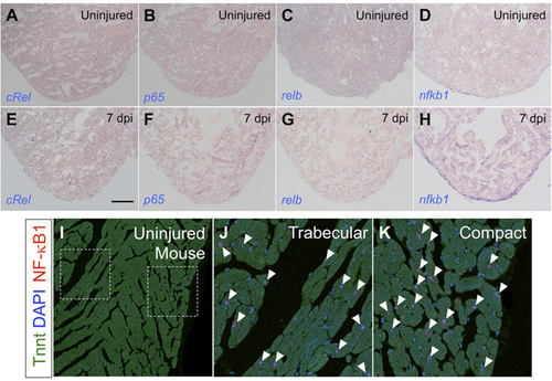
NF-KB factors during heart regeneration. (A–H) In situ hybridization for NF-KB transcription factors on sections from ventricles of cmlc2:CreER; βact2:RS-DTA animals treated with vehicle (A–D) or tamoxifen (E–H) to diffusely ablate cardiomyocytes. nfkb1 was the only gene from this group to show a detectable signal in cardiomyocytes. (I–K) Staining of uninjured adult mouse hearts for NF-KB1 and Tnnt, indicating that NF-KB1 is present in murine cardiomyocytes (arrowheads). We did not detect an obvious difference in NF-KB1+ cardiomyocytes in compact versus trabecular muscle. (Scale bar: 100 µm.) |
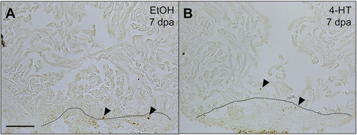
Cardiomyocyte apoptosis assays during NF-KB inhibition. TUNEL staining on sections of 7 dpa hearts from cmlc2:CreER; βact2:RS-IKBSR fish treated with vehicle (A) or 4-HT (B). No gross differences in TUNEL staining were noted during NF-KB inhibition. Arrowheads indicate TUNEL + nuclei. (Scale bar: 100 µm.) |
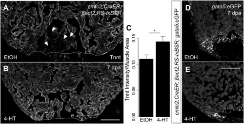
NF-KB signaling contributes to cardiomyocyte dedifferentiation. (A and B) Representative images of 7 dpa ventricles from cmlc2:CreER; βact2:RS-IKBSR treated with vehicle (n = 6) or 4-HT (n = 7) stained for Tnnt (gray). Arrowheads show diminished Tnnt staining in cardiomyocytes adjacent to the wound in control animals, suggestive of sarcomere disassembly. (C) Quantification of intensity of Tnnt intensity per muscle area suggests decreased sarcomere disassembly in ventricles with defective NF-KB signaling (n = 13). (D and E) Section images of 7 dpa ventricles from cmlc2:CreER; βact2:RS-IKBSR; gata5:eGFP fish treated with vehicle (n = 5) or 4-HT (n = 5). eGFP fluorescence is shown in grayscale. *P < 0.05, Student’s t test, two-tailed. (Scale bars: 100 µm.) |