
- Title
-
Expression patterns of CREB binding protein (CREBBP) and its methylated species during zebrafish development
- Authors
- Batut, J., Duboé, C., Vandel, L.
- Source
- Full text @ Int. J. Dev. Biol.
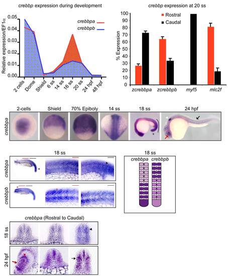
Expression patterns of crebbpa and crebbpb transcripts during zebrafish embryogenesis. Expression levels of crebbpa (red line) and crebbpb (blue line) were monitored by RT-qPCR at 2-cell, dome, shield, 6 ss, 14 ss, 16 ss, 20 ss, 24 hpf and 48 hpf. (B) Expression levels of crebbpa, crebbpb, myf5 and mlc2f in 10 anterior somites (rostral, red bars) and 10 posterior somites (caudal, black bars) of 20 ss embryos. Myf5 expression is specific of the nascent caudal somites while mlc2f, a specific marker of fast differentiating muscle fibres, is preferentially expressed in the rostral somites at this stage. All RT-qPCR data were normalized to EF1alpha housekeeping gene expression and data shown were from 3 independent experiments. (C) Whole-mount in situ hybridization against crebbpa at the indicated developmental stages. The line on the right at 18 ss indicates crebbpa enrichment in the trunk and caudal somites. *, PreSomitic Mesoderm; red arrow, lens; white arrowhead, otic vesicle; black arrow, somitic expression. (D) In situ hybridization against crebbpa and crebbpb at 18 ss (18 hpf) with rostral and caudal magnifications (red and black lines, respectively) of flat-mounted embryos. Black arrowhead, enrichment in caudal somites; red asterisk, enrichment in rostral somites. (E) Schematic summary of crebbpa and crebbpb expression at 18 ss. A dorsal view is represented (anterior to the top). (F) Transverse sections of 18 ss (upper panels) and 24 hpf (bottom panels) embryos from red (rostral) to black (caudal) lines noted in (D), showing crebbpa enrichment in the myotome at 18 ss (black arrowhead), in the lens (red arrow), in the retina (red arrowhead) and in the myotome (black arrow) at 24 hpf. |
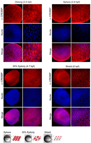
CREBBP protein expression patterns from blastula to gastrula. (A-D) Immunohistochemistry with CREBBP specific antibody at (A) oblong, (B) sphere, (C) 30% epiboly and (D) shield stages. Nuclei are visualized in blue with TO-PRO3 staining and a merged picture is shown. (E) Schematic illustration of CREBBP subcellular localization at these stages, cells are shown with CREBBP labeled in red. EXPRESSION / LABELING:
|
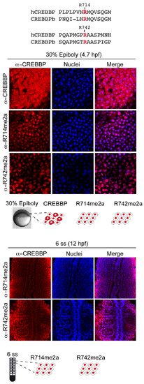
CREBBP-methylated species expression pattern at late blastula (30% epiboly) and during somitogenesis (6 ss). (A) Alignment of the epitopes of human CREBBP (hCREBBP) arginines R714 and R742 with the corresponding sequences of zebrafish crebbpa coding protein (CREBBPb or CBP-B). (B,C) Immunohistochemistry with CREBBP and CREBBPmethylated specific antibody as indicated at (B) 30% epiboly and (C) 6 ss. Nuclei are visualized in blue and a merged picture is shown. (B′, C′) Schematic illustration of subcellular localization of CREBBP and CREBBPmethylated proteins, with CREBBP labeled in red. EXPRESSION / LABELING:
|
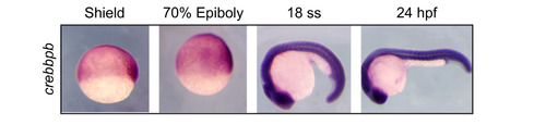
In situ hybridization against crebbpb at the indicated stages of development. The crebbpb transcript is ubiquitously expressed from gastrulation to 24 hpf. |
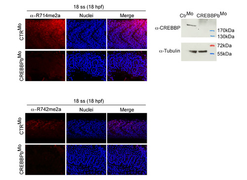
Validation of CREBBP-methylated specific antibodies on zebrafish embryos. (A,B) Immunohistochemistry with CREBBPmethylated specific antibody at 18 ss using (A) R714me2a or (B) R742me2a antibody on embryos injected with Control morpholino (CTRMo) and CREBBPb morpholino (CREBBPbMo). Nuclei are stained in blue with To-Pro3 and a merged picture is shown. Expression of CREBBP-R714me2a and CREBBP-R742me2a is strongly reduced in CREBBPbMo- as compared to controlMo-injected embryos. Injections were performed at the 1-cell stage with 6 ng of CTRMo or CREBBPbMo (Gene-tools). Morpholino sequences were: CREBBPb 5′-GACTGTTGCCACCTGCCATGCCCAT-3′ and Control 5′-CCTCTTACCTCAGTTACAATTTATA-3′. (C) Knock-down of CREBBPb reduces CREBBPb protein level in 14 ss old zebrafish embryos. Embryos injected with either CTRMo or CREBBPbMo were collected at 14 ss and processed for western blot analysis to detect CREBBP expression. Tubulin was used as a loading control and a protein ladder is shown. Whole cell extracts from 20 embryos were classically prepared in 40 ml Laemmli sample buffer. 5 ml of each sample (4 embryos) were loaded per lane and subjected to SDS-PAGE analysis. The following antibodies were used: anti-CREBBP (1/200, A22, santa Cruz), anti a-Tubulin (1:5000, Sigma, T9026), anti-Rabbit IgG, HRP conjugate (1:50000, Promega, W4011) and anti-Mouse IgG, HRP conjugate (1:10000, Promega, W4021). Protein Ladder, PageRuler prestained Protein Ladder (Fermentas, SM0671). EXPRESSION / LABELING:
PHENOTYPE:
|