
- Title
-
First Demonstration of Antigen Induced Cytokine Expression by CD4-1+ Lymphocytes in a Poikilotherm: Studies in Zebrafish (Danio rerio)
- Authors
- Yoon, S., Mitra, S., Wyse, C., Alnabulsi, A., Zou, J., Weerdenburg, E.M., van der Sar, A.M., Wang, D., Secombes, C.J., Bird, S.
- Source
- Full text @ PLoS One
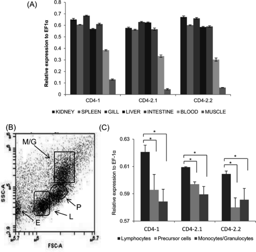
Zebrafish CD4-1/CD4-2 gene distribution. (A) Tissue/blood expression of CD4-1/CD4-2 genes in zebrafish. (B) Cell profile obtained from FACS sorted zf kidney and spleen cells. E = erythrocytes, L = lymphocytes, P = (hemopoietic) precursor cells and M/G = monocytes/granulocytes. (C) Differential expression of the zfCD4-1/CD4-2 genes within the FACS sorted populations. *, p<0.05. |

Zebrafish CD4-1 protein expression in transfected CHO cells and zebrafish tissues. (A) FACS analysis of zfCD4-1 positive cells in CHO cells transfected with zfCD4-1. Cells reacted with only secondary antibody or the pre-immune rabbit serum and secondary antibody are shown in the black and grey histogram, respectively, whilst cells incubated with zfCD4-1 polyclonal antibody and secondary antibody are shown in the red histogram. Note the increase in fluorescence intensity of the latter, as shown by a shift to the right. (B) and (C), Western blot analysis of zfCD4-1 protein. (B) Un-transfected CHO cell lysate (5 μl) (lane 1), and zfCD4-1 transfected CHO cell lysates (5μl, lane 2; 10μl, lane 3). (C), zf blood cell lysate (lane 1), kidney lysate (lane 2) and muscle lysate (lane 3). The data are representative of 3 independent experiments. |
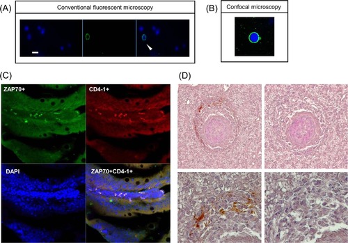
Detection of zfCD4-1+ cells using immunofluorescence assay and immuohistochemical assays. (A) Immunofluorescence staining of the zfCD4-1 molecule on zebrafish leukocytes. The cells stained with DAPI (blue) for counterstaining nuclei (left panel), the zfCD4-1 polyclonal followed by FITC labelled (green) secondary antibody (middle panel) and merged image (right panel) are shown. Scale bar = 10 µm). The arrow shows a cell expressing CD4 on its surface. (B) Cells from (A) were also visualized using a confocal microscope. (C) Double immunofluorescence staining of zebrafish peripheral lymphocytes in gut sections incubated with rabbit anti zfCD4-1 and mouse anti human ZAP70. ZAP70+ cells are green and CD4+ cells are red. Co-localization was confirmed by a Z-stack image analysis using Zeiss confocal microscopy. The data are representative results obtained from three independent experiments. (D) CD4-1+ cells present within the cuff of leukocytes surrounding the granulomas developed after M. marinum EspG5::Tn mutant infection of zebrafish 4 weeks earlier. Top images 200x, bottom images 400x. Left hand images used anti-CD4-1 serum, right hand images are controls. EXPRESSION / LABELING:
|