
- Title
-
Inexhaustible hair-cell regeneration in young and aged zebrafish
- Authors
- Pinto-Teixeira, F., Viader-Llargués, O., Torres-Mejía, E., Turan, M., González-Gualda, E., Pola-Morell, L., López-Schier, H.
- Source
- Full text @ Biol. Open
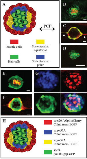
Neuromast organization and transgenic zebrafish lines. (A) Scheme of a neuromast depicting different cell types and axes of symmetry. (B-F) Confocal image of a larval neuromast of (B) Tg[Cldnb:mem-EGFP;ET(krt4:EGFP)sqet4] (green), incubated in the vital dye DiAsp to reveal functional hair cells (red), (C) Tg[Alpl:mCherry (red) ; ET(krt4:EGFP)sqet20 (green)] in which white arrowhead point to interneuromast cells, (D) Tg[pou4f3:gap43-GFP (green) ; pou4f3:Ribeye-Kusabira (red)], (E) Tg[ET(krt4:EGFP)sqgw57A] (green) labeled with DiAsp (red), (F) Tg[ET(krt4:EGFP)sqgw57A (green) ; Alpl:mCherry (red)], (G) Tg[ET(krt4:EGFP)sqgw57A] (green) immunostained for Sox-2 (red) and incubated with the nuclear dye DAPI (blue). (H) Overview of a neuromast with the different cell types and transgenic markers. Scale bars=10µm. |
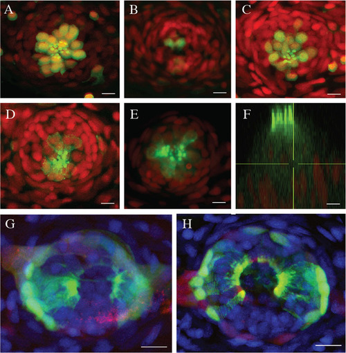
Efficient hair-cell regeneration in adult zebrafish. (A-C) Maximal projection of confocal images from Tg[ET(krt4:EGFP)sqet4] transgenics (green) showing neuromasts of the caudal fin of a 2-year old fish stained with DAPI (red) (A) before neomycin-treatment, (B) 2h after treatment, and (C) 72h after treatment. (D-E) Neuromast of an adult fish from Tg[myo6b:actb1-EGFP] transgenics (green) stained with DAPI (red) showing (D) hair cells controls, and (E) in neomycin-treated fish after hair-cell regeneration. (F) XZ profile of the same neuromast in E showing the apicobasal polarization of the regenerated hair cells (green). (G-H) Neuromast of an adult fish from Tg[Alpl:mCherry (red) ; ET(krt4:EGFP)sqet20 (green)] transgenics stained with DAPI (blue), showing (G) epithelial geometry in control fish and (H) in neomycin-treated fish at 72hpt. In all cases, N=8 neuromasts from 2 animals. Scale bars=10µm. |
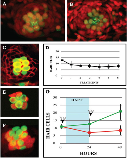
Hair-cell regeneration after recurrent damage. (A-B) Confocal images of a Tg[ET(krt4:EGFP)sqet4] (green) larval neuromast labeled with DAPI (red) showing hair cells (A) before neomycin treatment and (B) 72h after the 6th treatment. (C) Image of a Tg[ET(krt4:EGFP)sqet4] (green) larval neuromast counterstained for cellular membranes (red) 24hpt, with 8 hair cells. (D) Graph depicting the number of hair cells per neuromast 24h after each neomycin treatment, over the course of 6 consecutive treatments. (E-F) A neuromast from a neomycin-treated larva Tg[Atoh1a:dTomato (red) ; ET(krt4:EGFP)sqet4 (green)] without (E), and with Notch inhibition with DAPT (F), showing more numerous hair cells and stronger and broader Atho1a expression. (G) Graph showing number of hair cells per neuromast 24h after two neomycin treatments with (green) and without (red) inhibition of Notch. DAPT incubation period is shadowed in blue. Results are mean±s.d. Time points: 0h N=5 neuromasts (5 animals), 24h N=8 neuromasts (8 animals), 48h N=4 neuromasts (4 animals). |
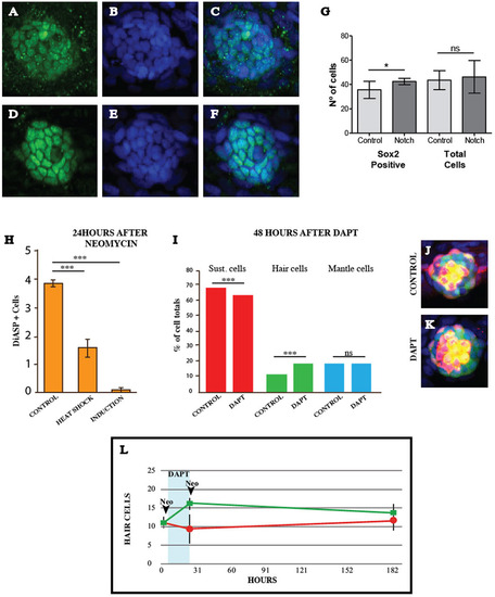
Neuromast organization after recurrent damage. (A-F) A neuromast immunostained for Sox-2 (red) and labeled with DAPI (blue) in (A-C) controls and (D-F) after induction of constitutive Notch signaling. (G) Quantification of Sox-2 positive cells and total cells number in regenerating neuromast 24h after neomycin treatment with and without induction of NICD in the double transgenic line Tg[Cldnb:Gal4ERT2;5xUAS-E1b:6xMYC-notch1a], results are mean±s.e.m. (Mann-Whitney test U=110, Control N=16 (3 larvae) and NIND=23 (4 larvae), *P=0.03). (H) Quantification of regenerated hair cells 24hpt after constitutive Notch activation by heat shock and chemical induction, results are mean±s.e.m. (Control versus Heat shock Mann-Whitney U=7.5, P<0.0001. Control versus Induction Mann-Whitney U=0, P<0.0001. Control N=33 (8 larvae), Heat shock N=7 (1 larvae), Induction N=14 (3 larvae) (I) Quantification of sustentacular, hair and mantle cells in regenerating neuromasts after two consecutive neomycin treatments, intercalated with a DAPT incubation period, results are mean±s.e.m. [Sust. cells Mann-Whitney U=61.5, P=0.0052. Hair cells Mann-Whitney U=34.5, P<0.0001. Mantle cells Mann-Whitney U=135, P=0.8081. Control N=22 (4 larvae), DAPT N=15 (3 larvae)]. (J-K) Representative neuromasts of the triple transgenic line Tg[ET(krt4:EGFP)sqgw57A ; Alpl:mCherry ; pou4f3: Kusabira-CAAX] used for quantifications. (L) Graph with hair-cell counts in regenerating neuromasts of Tg[ET(krt4:EGFP)sqet4] fish during 7days, showing controls (red) and DAPT-treated fish (green). DAPT incubation period is indicated by the blue vertical bar. Results are mean±s.d. Time points: 0h N=4 neuromasts (4 animals), 24h N=5 neuromasts (5 animals), 182h N=9 neuromasts (9 animals). |
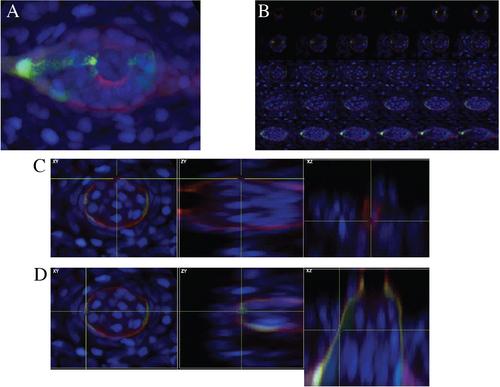
(A-D) A neuromast from an adult fish of the double transgenic line Tg[ET(krt4:EGFP)sqet20 (green); Alpl:mCherry (red)] counterstained with DAPI (blue), highlighting mantle cells (red) and the equatorial areas (green). (A) shows a maximal projection from an apical-to-basal Z-stack. (B) shows the series of individual stacks. (C) shows a single stack at a polar area, and (D) shows a single stack at an equatorial area. They reveal that Tg[ET(krt4:EGFP)sqet20] marks preferentially the equatorial areas. |
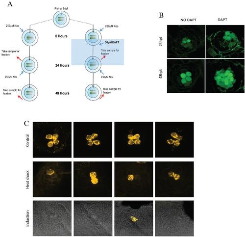
(A) A scheme showing the protocol for neomycin and DAPT treatments and hair-cell quantifications during regeneration. (B) representative neuromasts of Tg[ET(krt4:EGFP)sqet4 ; Cldnb:mem-EGFP], revealing hair cells 24 and 48 hpt, with and without DAPT treatments. (C) shows a series of neuromasts that reveal hair cells with DiASP labeling 24 hpt, after constitutive Notch activation by heat shock in the double transgenic line Tg[hsp70l:Gal4 ; 5xUAS-E1b:6xMYC-notch1a], and by chemical induction with tamoxifen in the transgenic line Tg[Cldnb:Gal4ERT2;5xUAS-E1b:6xMYC-notch1a]. |