
- Title
-
Dynamic visualization of transcription and RNA subcellular localization in zebrafish
- Authors
- Campbell, P.D., Chao, J.A., Singer, R.H., Marlow, F.L.
- Source
- Full text @ Development
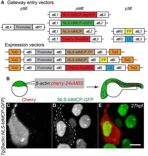
Transgenic zebrafish lines expressing NLS-tdMCP-eGFP can be used to detect transcripts in vivo. (A) Gateway compatible vectors for generation of NLS-tdMCP-FP and MS2-tagged RNAs. Plasmids used to generate transgenic lines by Tol2-mediated transgenesis are shown. 5′ Entry plasmids (p5E) containing the desired promoter elements can be recombined with NLS-tdMCP-eGFP or NLS-tdMCP-tagRFP entry plasmids (pME) for expression in any cell type or tissue. Similarly, the pME-NLS-tdMCP plasmid can be recombined with any in-frame fluorescent protein (FP) in a 3′ entry plasmid (p3E) to make custom NLS-tdMCP-FPs. RNA localization elements (LE) can be tagged with cherry-24xMBS by recombining the appropriate pME and p3E plasmids. (B) Schematic of the experiment used to validate in vivo MS2-MCP interactions. (C-E) Live imaging of embryos at the sphere stage shows that cytoplasmic puncta are visible only in cells expressing the Cherry reporter. In some cases, as shown in C, the Cherry reporter aggregates, but does not overlap with MCP-GFP cytoplasmic puncta, suggesting that this is independent of the MS2-MCP interaction. The dotted line denotes borders of cells expressing Cherry reporter. Scale bar: 10µm. |
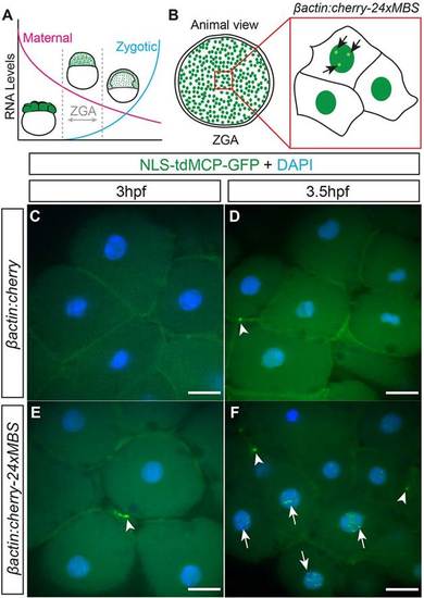
MS2-labeling reveals the onset of transcription in zebrafish embryos. (A) Immediately after fertilization, the RNA present is exclusively comprised of maternal products. After ZGA, zygotic transcripts begin to accumulate and replace maternal transcripts. (B) Embryos were injected with DNA encoding MS2-tagged-cherry RNA expressed from the βactin promoter and assayed for nuclear puncta around the time of ZGA (3-4.5hpf). (C-F) Animal pole view of fixed embryos showing that βactin:cherry-injected control embryos at (C) 3hpf and (D) 3.5hpf have no nuclear puncta. Animal pole views of fixed embryos injected with βactin:cherry-24xMBS at (E) 3hpf and (F) 3.5hpf showing that nuclear puncta (arrows) are not detected at 3hpf but are apparent at 3.5hpf and beyond. Both injected (C-F) and uninjected (supplementary material Fig. S2) embryos display accumulations of MCP-GFP at cell membranes (arrowheads). Scale bars: 25µm. |
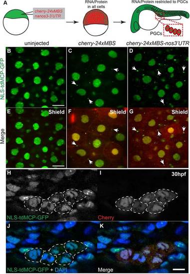
MS2-tagged nanos3 3′UTR is detected by MCP-GFP in the cytoplasm of PGCs. (A) Schematic depicting injection and imaging of cherry-24xMBS-nanos3 32UTR RNA. cherry-24xMBS-nanos3 3′UTR is initially in all cells but later is maintained only in PGCs. (B-G) Imaging of fixed embryos reveals strictly nuclear MCP-GFP in uninjected embryos at the shield stage (B,E) and cytoplasmic RNA visualized by MCP-GFP puncta in cherry-24xMBS- and cherry-24xMBS-nos3′UTR-injected embryos (C,D,F,G). Punctate accumulations of MCP-GFP are present on cell membranes of (F) cherry-24xMBS- and (G) cherry-24xMBS-nos3′UTR-injected embryos (arrows) indicating this represents a background artifact. Scale bars: 20µm. (H-K) The Cherry reporter reveals PGCs at 30hpf in embryos that were injected with cherry-24xMBS-nos32UTR. PGCs expressing Cherry display cytoplasmic MCP-GFP, whereas somatic cells and non-expressing PGCs have strictly nuclear MCP-GFP. The dotted lines denote borders of cells expressing Cherry reporter. Scale bar: 10µm. |
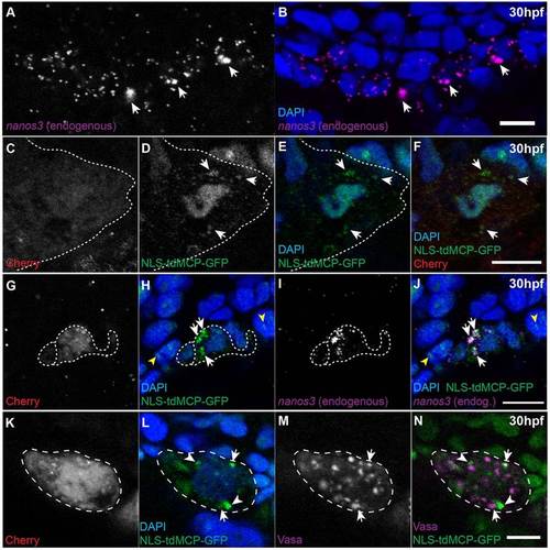
MS2-tagged nos3′utr colocalizes with endogenous nanos3 and a subset of Vasa-positive germ granules in PGCs. (A,B) Endogenous nanos3 RNA localizes to PGCs at 30hpf and is present throughout the cytoplasm and in larger perinuclear accumulations (arrows). Scale bar: 10µm. (C-F) Cherry-positive cells at 30hpf have diffuse cytoplasmic MCP-GFP signal and larger perinuclear accumulations (arrows). Scale bar: 10µm. (G-J) Accumulations of MCP-GFP in Cherry-expressing PGCs at 30hpf in cherry-24xMBS-nos3′UTR-injected embryos (white arrows) colocalize with endogenous nanos3 accumulations. Nonspecific nuclear aggregates of MCP-GFP (yellow arrowheads) do not colocalize with endogenous nanos3, consistent with these structures being artifacts. Scale bar: 25µm. (K-N) A subset of MCP-GFP accumulations in Cherry-expressing PGCs at 30hpf in cherry-24xMBS-nos3′UTR-injected embryos coincide with Vasa protein-positive germ granules (arrows) whereas others do not (arrowheads). Scale bar: 10µm. The dotted lines denote borders of cells expressing Cherry reporter. |