
- Title
-
Targeted germ line disruptions reveal general and species-specific roles for paralog group 1 hox genes in zebrafish
- Authors
- Weicksel, S.E., Gupta, A., Zannino, D.A., Wolfe, S.A., Sagerström, C.G.
- Source
- Full text @ BMC Dev. Biol.
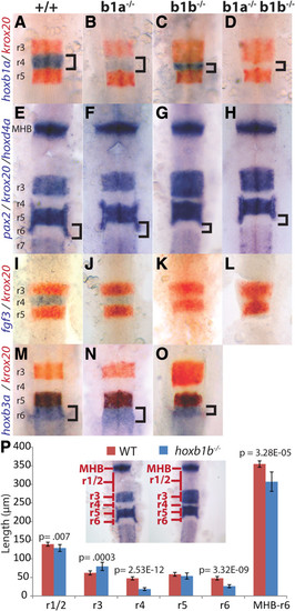
Zebrafish hoxb1b is required for hindbrain segmentation. A-O. Wild type (A, E, I, M), hoxb1a-/-(B, F, J, N), hoxb1b-/-(C, G, K, O) or doubly hoxb1a-/-;hoxb1b-/- embryos (D, H, L) were assayed by in situ hybridization for expression of hoxb1a in r4 (blue stain in A-D), krox20 in r3/r5 (red stain in A-D, I-O and blue stain in panels E-H), pax2 at the midbrain-hindbrain boundary (blue stain in E-H), fgf3 in r4 (blue stain in I-L), hoxd4a in r7 (blue stain in E-H) and hoxb3a in r5/r6 (blue stain in M-O). P. Quantification of segmentation defects in hoxb1b-/- embryos. Rhombomere lengths were measured as indicated in the inset. MHB-r6 measures the full distance from the anterior limit of the MHB to the posterior limit of r6. p-values were computed using Students’ t-test and error bars represent standard error. N = 10 embryos. All embryos are flat mounted in dorsal view with anterior to the top. A-H and M-O are at 22hpf, while I-L are at 14hpf. r = rhombomere; MHB = midbrain/hindbrain boundary. EXPRESSION / LABELING:
PHENOTYPE:
|
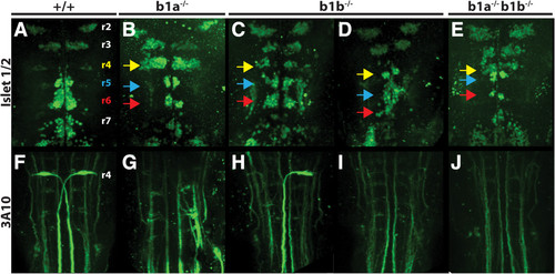
hoxb1a and hoxb1b are required for neuronal differentiation in the hindbrain. 48hpf wild type (A, F), hoxb1a-/-(B, G), hoxb1b-/-(C, D, H, I) or doubly hoxb1a-/-;hoxb1b-/- embryos (E, J) were assayed by immunostaining for the differentiation of branchiomotor neurons (islet1/2 staining in A-E) and Mauthner neurons (3A10 staining in F-J). Colored arrowheads indicate r4 (yellow), r5 (blue) and r6 (red). All embryos are flat mounted in dorsal view with anterior to the top. |
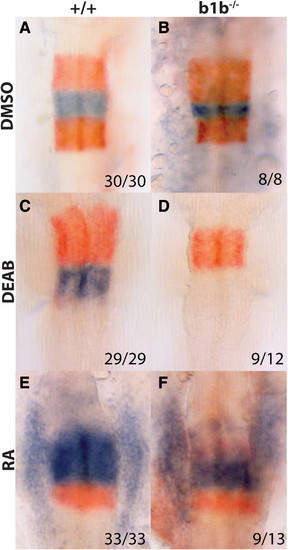
Retinoic acid and hoxb1b act independently to activate hoxb1a transcription. 19hpf wild type (A, C, E) or hoxb1b-/-(B, D, F) embryos were treated with DMSO (control; A, B), 10uM DEAB (C, D) or 100 nM RA (E, F) and assayed by in situ hybridization for expression of hoxb1a in r4 (blue stain in A-F) and krox20 in r3/r5 (red stain in A-F). All embryos are flat mounted in dorsal view with anterior to the top. EXPRESSION / LABELING:
|