
- Title
-
Discovery and Functional Annotation of SIX6 Variants in Primary Open-Angle Glaucoma
- Authors
- Carnes, M.U., Liu, Y.P., Allingham, R.R., Whigham, B.T., Havens, S., Garrett, M.E., Qiao, C., NEIGHBORHOOD Consortium Investigators, Katsanis, N., Wiggs, J.L., Pasquale, L.R., Ashley-Koch, A., Oh, E.C., Hauser, M.A.
- Source
- Full text @ PLoS Genet.
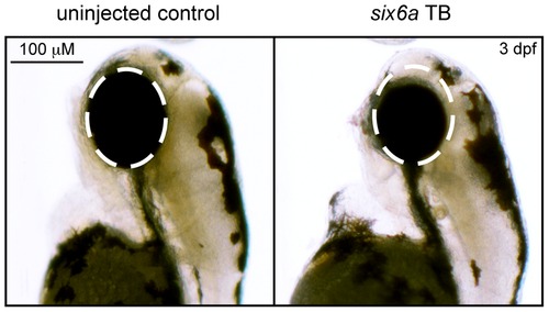
Morpholino knockdown of six6a. Zebrafish were microinjected with a six6a translation blocking morpholino. Lateral images, taken 3 days post fertilization (3 dpf), of a wild-type zebrafish (left) and a morpholino injected zebrafish (right) are shown, highlighting the small eye phenotype (dashed circle). PHENOTYPE:
|
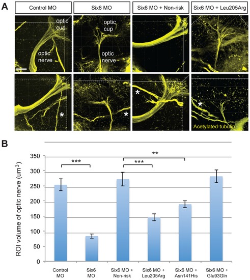
Functional evaluation of SIX6 variants on the volume of the optic nerve. Representative whole mount images of acetylated-tubulin expression in the heads of zebrafish embryos injected with a control or six6a morpholino, rescued by co-injection with human non-risk SIX6 transcript or a transcript containing the Leu205Arg hypomorphic variant (A). Acetylated-tubulin staining is restricted primarily to axon tracts and can be used to visualize the optic nerve. Relative to the control morphants, volumetric regions of interest (ROI) along the optic nerve in six6a morphants were reduced significantly. Co-injection of human variants revealed a hypomorphic (Leu205Arg, Asn141His) or benign (Glu93Gln) role of the variants on the optic nerve (B). Sample size for all injection paradigms ranged from 7–9 and p-values are plotted for each comparison (*** p<0.001; ** p<0.01). No significant changes in the volume of other axonal tracts in the head (marked by an asterisk) were detected. Standard error of the mean is shown and white scale bars = 20 μm. |
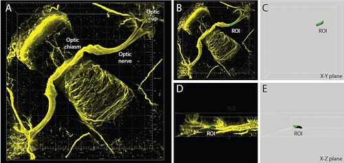
Volumetric analysis of the optic nerve. Confocal image of a 2 dpf zebrafish head stained with antibody to acetylated tubulin to visualize axons. (A) Regions of Interest (ROIs) 7.5 μm×7.5 μm×15 μm in size along the optic nerve were selected from reconstructed 3-dimensional images using Imaris software [X–Y plane (B–C) and X–Z plane (D–E)]. Panels C and E show the reconstructed ROI from which volumetric measurements are calculated. EXPRESSION / LABELING:
|