
- Title
-
Activating mutations in STIM1 and ORAI1 cause overlapping syndromes of tubular myopathy and congenital miosis
- Authors
- Nesin, V., Wiley, G., Kousi, M., Ong, E.C., Lehmann, T., Nicholl, D.J., Suri, M., Shahrizaila, N., Katsanis, N., Gaffney, P.M., Wierenga, K.J., Tsiokas, L.
- Source
- Full text @ Proc. Natl. Acad. Sci. USA
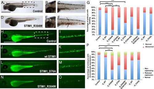
Expression of STIM1_R304W, but not ORAI1_P245L, results in bleeding and reduced expression and defective flow of thrombocyte progenitors in zebrafish embryos. (A–F) Whole-body lateral views of a control (A) and STIM1_R304W (D) injected embryos at 48 h post fertilization stained with o-dianisidine. B and E are magnifications of the cephalic boxed area in A in all respective embryos, showing brain hemorrhages (white arrows) in the STIM1_R304W mRNA injected embryos (E). Panels C and F show magnified views of the boxed caudal area in A in the respective conditions. Areas of intrasomitic and caudal bleeding are highlighted with black arrows. (G) Percent distribution of normal embryos vs. embryos with spontaneous bleeding episodes (**P < 0.001, ***P < 0.0001). (Scale bar: 500 µm.) Whole-body lateral views of control (H), STIM1_WT- (J), STIM1_D76A- (L), and STIM1_R304W- (N) injected Tg(CD41:GFP) embryos at 72 h post fertilization. I, K, M, and O are magnifications of the ventral boxed area in H in the respective embryos, showing expression of GFP in thrombocytes. The boxed area corresponds to the site where early hematopoiesis takes place in zebrafish. (P) Percent distribution of normal embryos vs. embryos with reduced or no expression of thrombocyte progenitors (*P < 0.05, **P < 0.001, and ***P < 0.0001). EXPRESSION / LABELING:
|

Overexpression of STIM1_R304W causes hypoplastic caudal vein formation in developing zebrafish embryos. (A–D) Lateral views of a control (A) or STIM1_R304W (C)-injected Tg(Fli1:GFP) transgenic embryo expressing GFP in endothelial cells and hence allowing the visualization of developing blood vessels 48 hpf. B and D are magnified views of the boxed area in A in the respective embryos, showing that the caudal vein in the STIM1_R304W injected embryos (C) appears hypoplastic compared with the controls (B). (E) Percent distribution of normal vs. abnormal embryos as determined by phenotypic aberrations of the caudal vein (**P < 0.001, ***P < 0.0001). EXPRESSION / LABELING:
|