
- Title
-
Zebrafish yap1 plays a role in differentiation of hair cells in posterior lateral line
- Authors
- Loh, S.L., Teh, C., Muller, J., Guccione, E., Hong, W., Korzh, V.
- Source
- Full text @ Sci. Rep.
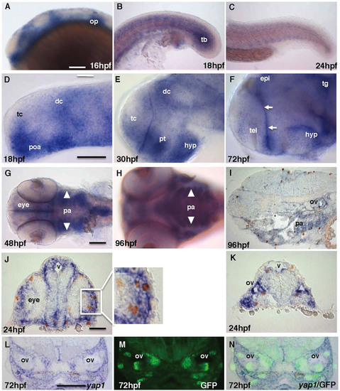
(A–C) yap1 is expressed in the brain, somites and tail bud until mid-somitogenesis and its expression in the posterior body declines later on. (D–F) yap1 expression become more restricted to the ventricular zones (proliferative region, arrows) in the brain as the embryo matures. (G–I) Staining of yap1 in pharyngeal arch (arrowhead) at 48–96 hpf. (J), (K) Most proliferative cells detected by anti-pH3 antibody (brown, cell nucleus) are yap1-positive (blue) in cross-section of 24 hpf and (I) sagittal-section of 96 hpf embryos. L-N – cross-sections at the mid-hindbrain (ear) level of 72 hpf SqET33-mi60A transgenic larvae (lnfg, progenitors of sensory cells). (L) yap1 in situ hybridization; (M) GFP expression; (N) composite yap1 in situ hybridization/GFP expression. (A–F) – lateral view, (G), (H) – ventral view. Scale bar = 40 μm. Abbreviations: dc, diencephalon; epi, epiphysis; hyp, hypothalamus; op, otic placode; pa, pharyngeal arches; poa, preoptic area; pt, prethalamus; tb, tail bud; tc, telencephalon; tg, tegmentum. EXPRESSION / LABELING:
|
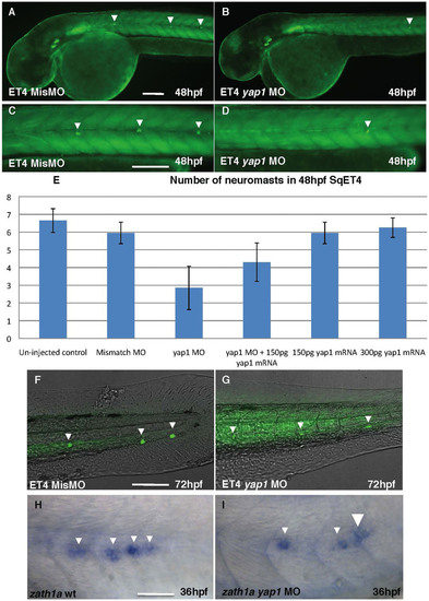
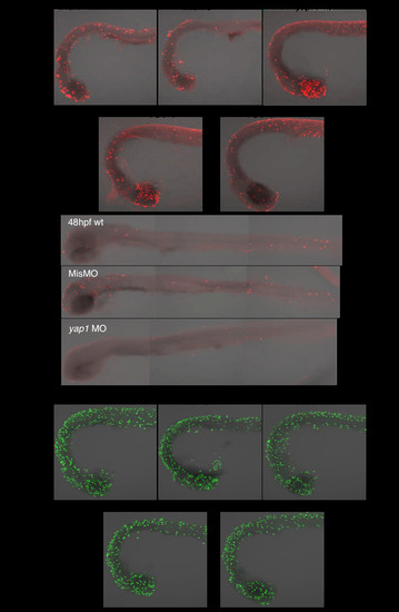
(A), (C), (F) – controls; (B), (D), (G) – Yap1 morphants. All embryos are shown in lateral view. The table illustrates the rescue effect of yap1 mRNA coinjection. The original data are provided as a supplement. (A), (B) – head and anterior trunk, (C), (D) – intermediate trunk, (F), (G) – tail. Notice a reduction in a number of neuromast cells and increase of background due to longer exposure in (G) comparing to (F). (H), (I) – zath1a expression in the lateral line primordium of controls (H) and morphants (I). Scale bar = 40 μm, (H)&(J) = 20 μm. EXPRESSION / LABELING:
PHENOTYPE:
|
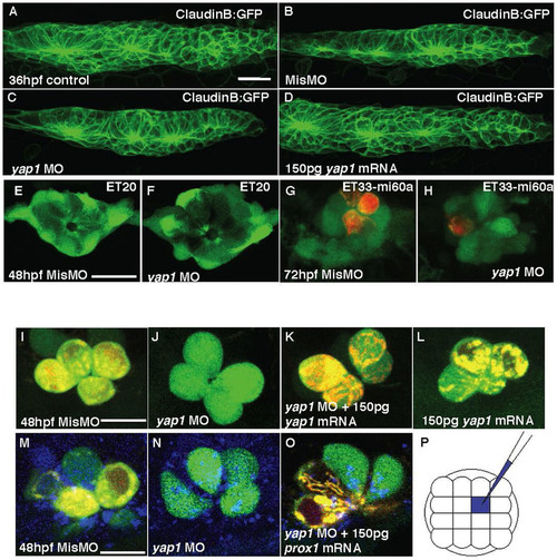
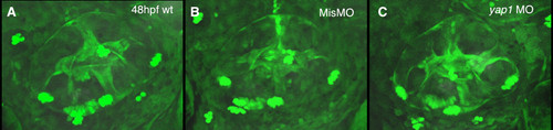
(A–D) Primordium I (PrimI) in yap1 morphants. (E) Size of PrimI at 36 hpf. (E, F) Mantle cells of neuromast in 72 hpf ET20 yap1 morphants. (G–H) Mechanoreceptors of neuromast (red, DiAsp) in 72 hpf ET33-mi60a embryos, support cells (green). (I–L) hair cells. (J–L) Loss of yap1 abolished the functionality of hair cells in SqET4 transgenics, stained with DiAsp (red); it was rescued with yap1 mRNA injection. (M–O) Mismatch MO, yap1 MO or yap1 MO with prox1 mRNA were co-injected with a marker (Cascade Blue) into single cell of SqET4 embryos at 16-cell stage. (P) The schematic drawing of injected single cell (blue), any one of the middle four cells, at 16-cell stage. Scale bar = 10 μm. EXPRESSION / LABELING:
|
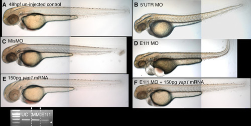
yap1 morphants could be partially rescued with yap1 mRNA. (A) wild type controls and (C) mismatched MO, (B) Translational (5′UTR MO), and (D) splice-site (E1I1 MO) morphants, co-injected with p53 MO, have similar phenotype; curved up trunk, cardiac edema, reduced head and eye size (E) Overexpression of Yap1 caused no obvious phenotype. (F) Partial rescue was achieved by co-injection of E1I1 MO with 150pg of yap1 mRNA. (G) Band-shift of yap1 mRNA transcript (arrow) is observed in RT-PCR of E1I1 morphants (E1I1) as compared to un-injected control (UC) and mismatch control (MM). |
Knockdown of yap1 increased apoptosis. (A) TUNEL-positive cells (red) were increased in yap1 morphant mostly in the head, which could be partially rescued with co-injection of yap1 mRNA. (B) No significant cell death was observed in posterior lateral line system of yap1 morphants at 48hpf. (C) Cell proliferation in both gain and loss-of-function of yap1 was not obviously different from that in controls (anti-pH3 antibody staining, green). |
Otic vesicle. The otic vesicle of morphants appears somewhat reduced, but its patterning is relatively normal. |