
- Title
-
Inter-cellular exchange of cellular components via VE-cadherin-dependent trans-endocytosis
- Authors
- Sakurai, T., Woolls, M.J., Jin, S.W., Murakami, M., Simons, M.
- Source
- Full text @ PLoS One
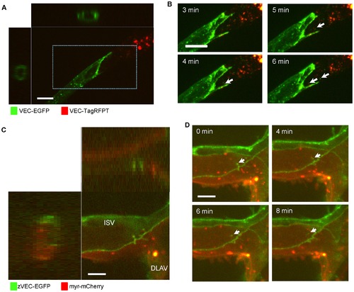
VEC trans-endocytosis occurs in sprouting HUVECs and endothelial cells in zebrafish embryos. (A) A single z-plane, x-z and y-z cross-sectional images of the tube-like structure of sprouting HUVECs. HUVECs expressing VEC-EGFP and HUVECs expressing VEC-TagRFPT were co-cultured in three-dimension fibrin gels. The Z-stack images were taken at the connection between HUVECs expressing VEC-EGFP and HUVECs expressing VEC-TagRFPT. Images were collected at 0.3 μm intervals with the 488 nm and 561 nm lasers to create a stack in the Z axis with a 60x objective. Scale bar = 20 μm. See also Movie S5. (B) Higher magnification of a 3-dimensional projection image of the indicated area in A. Arrows show VEC-EGFP molecules were trans-endocytosed by HUVECs expressing VEC-TagRFPT. Scale bar = 10 µm. (C) A three dimensional projection image, x-z and y-z cross-sectional images of the connection between the dorsal longitudinal anastomotic vessel (DLAV) and an intersegmental vessel (ISV) of a zebrafish embryo. Zebrafish VEC-EGFP (zVEC-EGFP) plasmids were injected into Tg(flkl:myr-mCherry) zebrafish using Tol2 system for transient mosaic expression of zVEC-EGFP. Images were collected at 1 µm intervals using the 488 nm and 561 nm lasers to create a stack in the Z axis with a 60x objective. Scale bar = 5 μm. See also Movie S6. (D) Sequential 3-dimensional projection image of the zebrafish vessel in C. Arrows show a zVEC-EGFP positive structure budding to inside of the endothelial cell. Scale bars = 5 μm. |