
- Title
-
The role of inab in axon morphology of an identified zebrafish motoneuron
- Authors
- Van Ryswyk, L., Simonson, L., and Eisen, J.S.
- Source
- Full text @ PLoS One
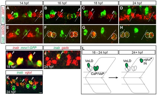
inab is dynamically expressed in PMNs and INs. (A–K) Single confocal slices of embryos labeled with inab, islet, and neurotransmitter riboprobes (gads for GABA and vglut for glutamate). Diagonal lines represent somite boundaries. At 14 hpf, inab is coexpressed with neither islet1 (A) nor islet2a (E). Between 16 hpf and 24 hpf, inab is expressed in islet2a+ PMNs (circled cells in F–H) but not islet1+ PMNs (arrowheads in A–C). inab is expressed in the VeLD IN, as determined by coexpression with both GFP in mnx1:GFP transgenic embryos (arrowheads, I) and gad mRNA (arrowhead, J). At 24 hpf, inab is coexpressed with vglut (arrowheads, K) in a cell dorsal to the VeLD IN (circle, K). (L) Schematic of inab mRNA dynamics during early development. Between 16–24 hpf, inab is expressed in both CaP and VaP MNs and in VeLD INs. After 24 hpf, inab expression in CaP and VaP is downregulated, although it persists in VeLD and an additional, dorsally-located, glutamatergic IN. Scale bar, 5 μm in A–J; 10 μm in K. EXPRESSION / LABELING:
|
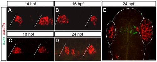
inaa is expressed in the zebrafish head and not in PMNs. (A–E) Z-projections of confocal stacks of embryos labeled with inaa and islet2a riboprobes. Diagonal lines represent somite boundaries. Between 14 hpf and 24 hpf, inaa is never coexpressed with islet2a in the trunk (A–D). At 24 hpf, inaa expression can be seen in a region of the head that corresponds to the approximate location of the diencephalic ventricle (E). Boundaries of the fish head and eyes are marked with a dotted line. Scale bar 10 μm in A–D; 50 μm in E. EXPRESSION / LABELING:
|
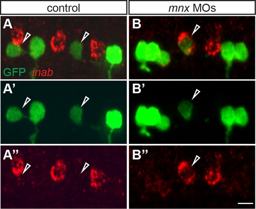
inab is expressed in MiP-CaP hybrids generated by Mnx protein knockdown. (A–B) Z-projections of confocal stacks of control and Mnx MO-injected nrp1a:GFP embryos labeled with inab riboprobe and anti-GFP antibody. At 26 hpf, inab is not expressed in GFP+ MiP MNs in control embryos (arrowheads, A-A3), but is expressed in a small percentage of GFP+ MiP MNs in MO-injected embryos (arrowhead, B-B3). Scale bar, 10 μm. EXPRESSION / LABELING:
|
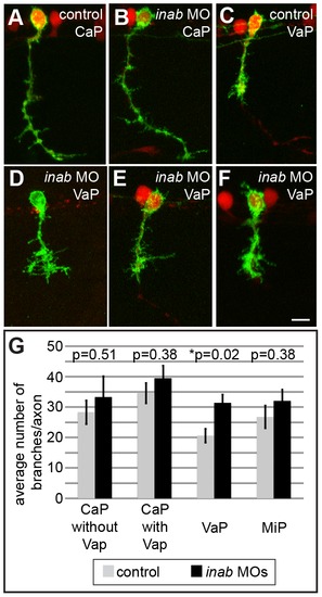
inab is required for morphology of VaP axons. (A–F) Z-projections of confocal images of control and inab MO-injected embryos. At 26 hpf, UAS:EGFP plasmid injection into mnx1:GAL4;UAS:tdTomato transgenic embryos reveals similar overall morphology of CaP neurons in both control (A) and MO-injected embryos (B). VaP axons in MO-injected embryos (D–F) have more processes than VaP axons in control embryos (C). (G) Graph representing average number of branch points per axon in individually-labeled cells in control and MO-injected embryos. No significant difference between conditions for CaP neurons alone (n = 11, p = 0.51), CaP MNs that are located next to VaP MNs (n = 11, p = 0.38), or MiP MNs (n = 10, p = 0.38). Only the increase in VaP axon processes is significant (n = 14, p = 0.02). Scale bar is 10 μm in A–F. PHENOTYPE:
|
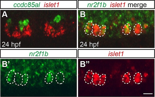
ccdc85al and nr2f1b are expressed in the zebrafish spinal cord. (A–B) Single confocal slices of 24 hpf embryos labeled with riboprobe. ccdc85al is expressed in cells just dorsal to the islet1+ MNs (A). nr2f1b is expressed broadly throughout the spinal cord, including expression in islet1+ MNs (circles, B-B3). Scale bar, 10 μm. EXPRESSION / LABELING:
|
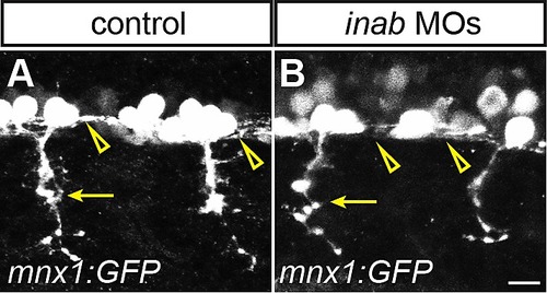
inab knockdown does not affect ventral motor nerve or VeLD axons. (A–B) Z-projections of confocal images of control and inab MO-injected mnx1:GFP transgenic embryos. At 20 hpf, both ventral CaP axons (arrows, A–B) and descending VeLD axons (arrowheads, A–B) are visible in both control (A) and inab MO-injected (B) embryos. Scale bar, 10 μm. EXPRESSION / LABELING:
|