
- Title
-
Expression patterns of dnmt3aa, dnmt3ab, and dnmt4 during development and fin regeneration in zebrafish
- Authors
- Takayama, K., Shimoda, N., Takanaga, S., Hozumi, S., and Kikuchi, Y.
- Source
- Full text @ Gene Expr. Patterns
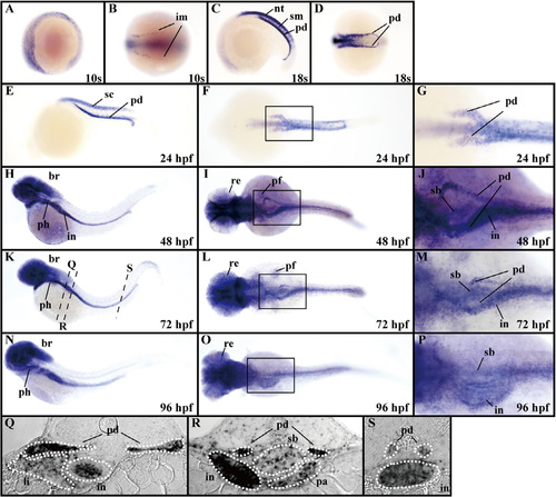
Expression patterns of dnmt3aa in zebrafish embryos and larvae. (A–P) dnmt3aa expression was examined by whole-mount in situ hybridization at the 10s and 18s stages as well as at 24, 48, 72, and 96 hpf. Lateral views, anterior to the left (A, C, E, H, K, and N). Vegetal pole views of the tail-bud region, anterior to the left (B and D). Dorsal views, anterior to the left (F, I, L, and O). The boxed areas in (F), (I), (L), and (O) are shown enlarged in (G), (J), (M), and (P), respectively. (Q–S) Transverse sections showing dnmt3aa expression. The transverse sections were cut at the levels indicated by the dashed black lines in (K). br, brain; im, intermediate mesoderm; in, intestine; li, liver; nt, neural tube; pa, pancreas; pd, pronephric duct; pf, pectoral fin buds; ph, pharyngeal arches; re, retina; sb, swim bladder; sc, spinal cord; sm, somitic mesoderm. EXPRESSION / LABELING:
|
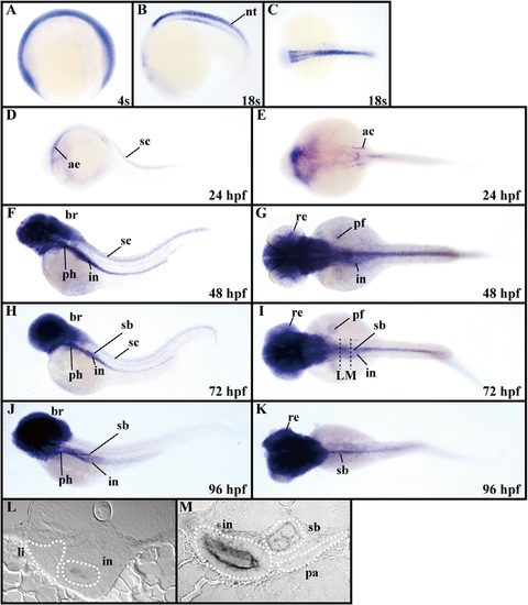
Expression patterns of dnmt3ab in zebrafish embryos and larvae. (A–K) dnmt3ab expression was examined by whole-mount in situ hybridization at the 4s and 18s stages, and at 24, 48, 72, and 96 hpf. Lateral views, anterior to the left (A, B, D, F, H, and J). Dorsal views, anterior to the left (C, E, G, I, and K). (L and M) Transverse sections showing dnmt3ab expression. The transverse sections were cut at the levels indicated by the dashed black lines in (I). ac, auditory capsule; ae, anterior endoderm; br, brain; in, intestine; nt, neural tube; li, liver; pa, pancreas; pf, pectoral fin buds; ph, pharyngeal arches; re, retina; sb, swim bladder; sc, spinal cord. EXPRESSION / LABELING:
|
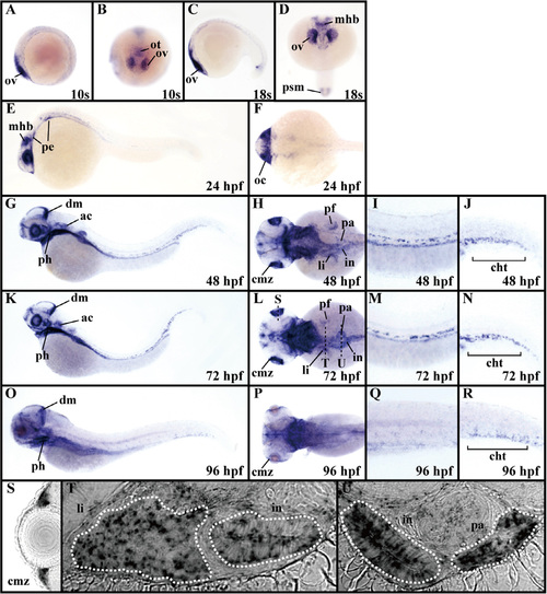
Expression patterns of dnmt4 in zebrafish embryos and larvae. (A–R) dnmt4 expression was examined by whole-mount in situ hybridization at the 10s and 18s stages as well as at 24, 48, 72, and 96 hpf. Lateral views, anterior to the left (A, C, E, G, K, and O). Dorsal views, anterior to the bottom (B and D). Dorsal views, anterior to the left (F, H, L, and P). Magnified views of the agm (I, M, and Q) and cht (J, N, and R) in (G), (K), and (O). (S–U) Cross and transverse sections showing dnmt4 expression at 72 hpf. The cross and transverse sections were cut at the level indicated by the dashed black lines in (L). ac, auditory capsule; cht, caudal hematopoietic tissue; cmz, ciliary marginal zone; dm, dorsal midbrain; in, intestine; li, liver; mhb, midbrain-hindbrain boundary; oc, optic cup; ot, optic tectum; ov; optic vesicle; pa, pancreas; pe, pharyngeal endoderm; pf, pectoral fin buds; ph, pharyngeal arches; psm, presomitic mesoderm. EXPRESSION / LABELING:
|
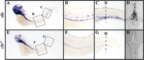
dnmt4 is expressed in HSCs/hematopietic cells in agm and cht in zebrafish larvae. (A–H) dnmt4 expression in sibling and clos5 mutant larvea was examined by wholemount in situ hybrididnmt4 is expressed in HSCs/hematopietic cells in agm and cht in zebrafish larvae. (A–H) dnmt4 expression in sibling and clos5 mutant larvea was examined by whole-mount in situ hybridization at 72 hpf. Lateral views, anterior to the left (A–C, and E–G). Magnified views of the agm (B and F) and cht (C and G) in (A) and (E). (D and H) Transverse sections showing dnmt4 expression. The transverse sections were cut at the levels indicated by the dashed black lines in (C) and (G). clos5, cloches5; sib, sibling. Transverse sections showing dnmt4 expression. The transverse sections were cut at the levels indicated by the dashed black lines in (C) and (G). clos5, cloches5; sib, sibling. EXPRESSION / LABELING:
PHENOTYPE:
|
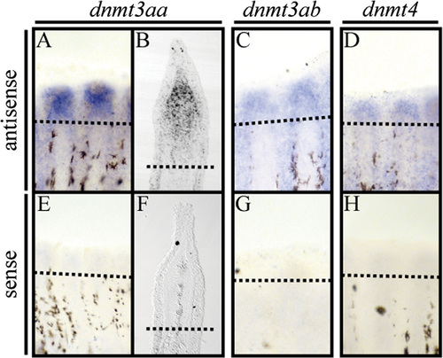
Expression patterns of dnmt3aa, dnmt3ab, and dnmt4 during zebrafish fin regeneration. (A–H) In situ hybridization analyses with antisense riboprobes (A–D) and sense riboprobes (E–H) of dnmt3aa, dnmt3ab, and dnmt4 in fin regenerates at 72 hpa. Longitudinal sections of wildtype fin regenerates (B and F). Dashed black lines indicate the amputation planes. EXPRESSION / LABELING:
|

Expression of dnmt4 in the brain at 72 hpf. (A–E) dnmt4 and sonic hedgehog a (shha) expressions were examined by whole-mount in situ hybridization at 72 hpf. Lateral views, anterior to the left (A and D). Magnified views of sagittal sections of dnmt4 expression (B and C) and shha expression (E) in (A) and (D), respectively. Brain anatomy and comparison with shha expression show that dnmt4 is expressed in the te, dt, vt, and p. ce; cerebellum; dt, dorsal thalamus; p, pallium; te, tectum; vt, ventral thalamus; zli, zona limitans intrathalamica. EXPRESSION / LABELING:
|
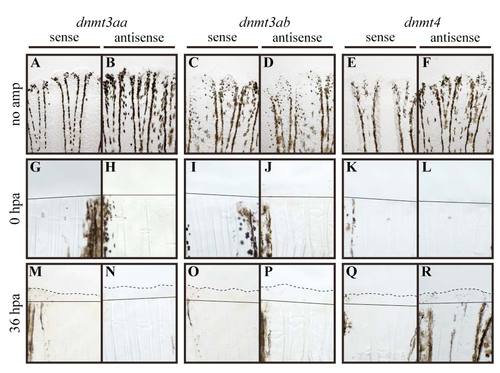
Expression patterns of dnmt3aa, dnmt3ab, and dnmt4 in adult fin, immediately after fin amputation, and at 36 hpa. (A–R) in situ hybridization analyses with antisense riboprobes (B, D, F, H, J, L, N, P, and R) and sense riboprobes (A, C, E, G, I, K, M, O, and Q) of dnmt3aa, dnmt3ab, and dnmt4 before (no amp) and immediately after fin amputation (0 hpa), and at 36 hpa. Black lines and dashed black lines indicate the amputation planes and the margins of the blastema, respectively. no amp, no amputation. |
Reprinted from Gene expression patterns : GEP, 14(2), Takayama, K., Shimoda, N., Takanaga, S., Hozumi, S., and Kikuchi, Y., Expression patterns of dnmt3aa, dnmt3ab, and dnmt4 during development and fin regeneration in zebrafish, 105-110, Copyright (2014) with permission from Elsevier. Full text @ Gene Expr. Patterns