- Title
-
Bosutinib inhibits migration and invasion via ack1 in kras mutant non-small cell lung cancer
- Authors
- Tan, D.S., Haaland, B., Gan, J.M., Tham, S.C., Sinha, I., Tan, E.H., Lim, K.H., Takano, A., Krisna, S.S., Thu, M.M., Liew, H.P., Ullrich, A., Lim, W.T., and Chua, B.T.
- Source
- Full text @ Mol. Cancer
|
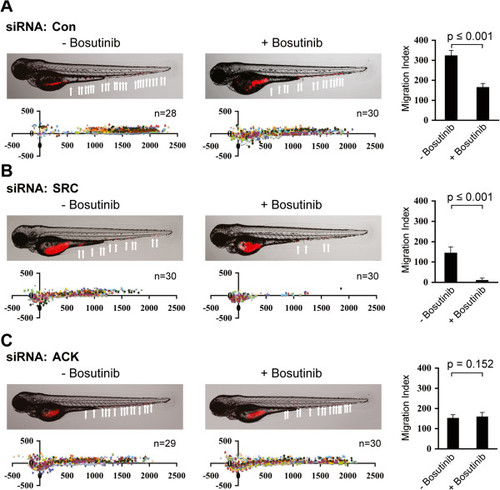
Reduction of lung cancer cell metastasis in zebrafish embryos by bosutinib is ACK1 dependent. Embryo injected with Vibrant DiD labeled NCI-H2009 cells showing tumor foci burden determined by segmented red channel (upper) and scatter plot representation of cell dissemination (bottom). Each data series represents one fish and number of injected embryos from 3 biological replicates is indicated (n). Comparative inhibition of cell migration, as measured by Migration Index, between untreated and drug pretreated cells is represented as bar graph (right), Mean ± SD. (A) Embryos injected with NCI-H2009 transfected with control siRNA. (B) Embryos injected with NCI-H2009 transfected with SRC siRNA. (C) Embryos injected with NCI-H2009 transfected with ACK1 siRNA. |
|
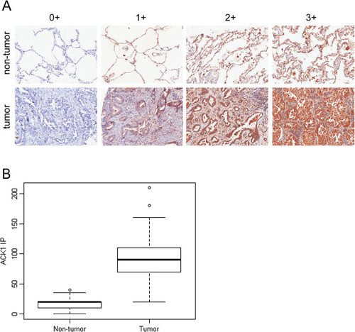
ACK1 is highly expressed in lung adenocarcinoma. Immunohistochemistry staining was performed on 210 NSCLC tumor and paired non-tumor sections on an in-house TMA using anti-ACK1 (C20, see material and method). (A) ACK1 immuno-positivity was defined as presence of brown cytoplasmic staining. Staining intensity was scored as 0, 1+, 2+ and 3+ (no, weak, moderate and strong staining, respectively). (B) Percentage of positively stained tumor cells was assessed as proportion of total number of tumor cells present in the section. Intensity percentage score, IP, was defined as product of the maximum immunostaining intensity and percentage of tumor cells stained. |


