
- Title
-
Whole genome sequencing in patients with retinitis pigmentosa reveals pathogenic DNA structural changes and NEK2 as a new disease gene
- Authors
- Nishiguchi, K.M., Tearle, R.G., Liu, Y.P., Oh, E.C., Miyake, N., Benaglio, P., Harper, S., Koskiniemi-Kuendig, H., Venturini, G., Sharon, D., Koenekoop, R.K., Nakamura, M., Kondo, M., Ueno, S., Yasuma, T.R., Beckmann, J.S., Ikegawa, S., Matsumoto, N., Terasaki, H., Berson, E.L., Katsanis, N., and Rivolta, C.
- Source
- Full text @ Proc. Natl. Acad. Sci. USA
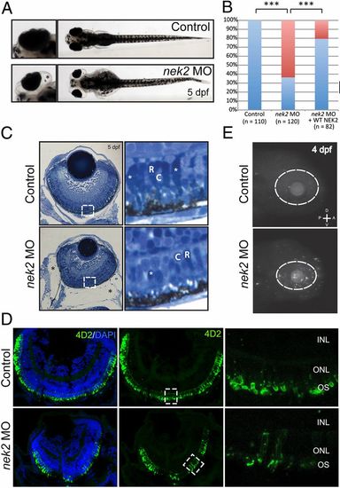
In vivo functional evaluation of nek2 loss in zebrafish. (A) Bright-field representation of 5-dpf control and nek2 morphant zebrafish embryos. Magnified Insets highlight ocular phenotypes including microphthalmia and enlarged eye sockets (marked by the black asterisk). (B) Ocular phenotypes including microphthalmia and enlarged eye sockets vs. normal phenotypes (red bars and blue bars, respectively) are quantified in control and nek2 morphant embryos, as well as in morphant animals rescued with human WT NEK2 mRNA. Asterisks indicate statistically significant differences between groups (P < 0.001). (C) Histology of control and nek2 morphant embryos also show enlarged eye sockets (marked by black asterisks) and microphthalmia. Magnified Insets show a decrease in the number of photoreceptors with apparent changes in domains of condensed chromatin (white asterisks). C, cones; R, rods. (D) Immunohistochemical analyses of retinal cryosections from control and nek2 MO embryos, stained with DAPI (blue) and the 4D2 antibody against rhodopsin (green). Suppression of nek2 results in the depletion of rods and in the mislocalization of rod opsin from the outer segment (OS) of photoreceptors. INL, inner nuclear layer; ONL, outer nuclear layer. (E) TUNEL immunofluorescent images of 4-dpf embryos, showing an increase in the number of apoptotic cells in nek2 morphant embryos. The dotted ovals indicate the position of the eye. A, anterior; D, dorsal; P, posterior; V, ventral. |
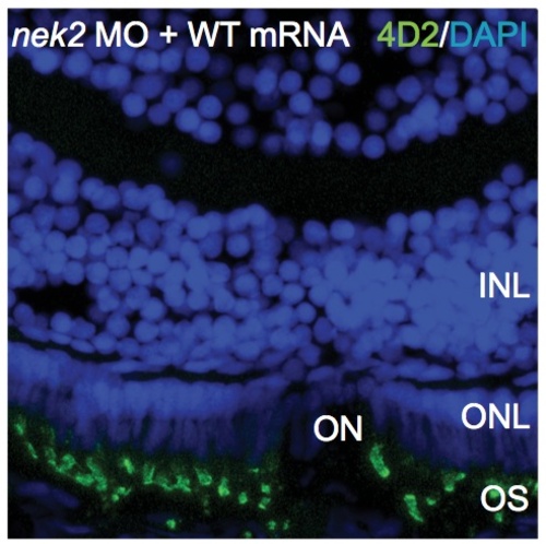
In vivo rescue of nek2 deficiency in zebrafish by human NEK2 mRNA supplementation. Immunohistochemical analyses of retinal cryosections from nek2 MO embryos + human wt NEK2 mRNA, stained with DAPI (blue) and 4D2 antibody (green). INL, inner nuclear layer; ONL, outer nuclear layer; ON, optic nerve; OS, outer segment of photoreceptors. |

In vivo functional evaluation of nek2 and rpgr deficiency in zebrafish. (A) Ocular phenotypes are quantified in control, nek2 MO, rpgr MO, and nek2 MO + rpgr MO embryos at full and sub-effective doses. (B) Immunohistochemical analyses of retinal cryosections from control, nek2 MO, rpgr MO, and nek2 + rpgr MO embryos at sub- effective doses, stained with DAPI (blue) and 4D2 antibody (green). Asterisk denotes excessive rhodopsin in the outer nuclear layer (ONL). OS, outer segment of photoreceptors; INL, inner nuclear layer. PHENOTYPE:
|