
- Title
-
Planar cell polarity proteins differentially regulate extracellular matrix organization and assembly during zebrafish gastrulation
- Authors
- Dohn, M.R., Mundell, N.A., Sawyer, L.M., Dunlap, J.A., and Jessen, J.R.
- Source
- Full text @ Dev. Biol.
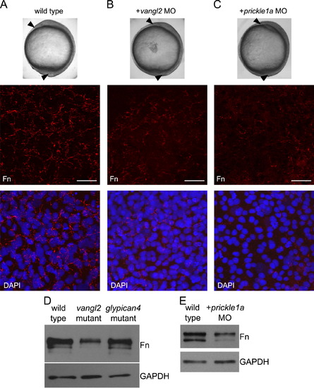
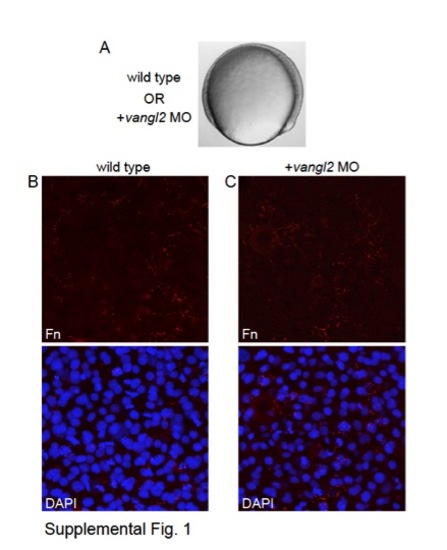
Knockdown of vangl2 and prickle1a decreases fibronectin. (A–C) Top panels show morphological convergence and extension phenotypes at tailbud stage (see arrowheads) of wild type, vangl2 morphant, and prickle1a morphant embryos. Middle/bottom panels show confocal images of fibronectin (Fn) immunolabeling without and with nuclear DAPI staining. Scale bars=20 μm. (D and E) Fn and GAPDH western blots of 10 μg total protein extract obtained from tailbud stage wild type, vangl2 mutant, glypican4 mutant, and prickle1a morphant embryos. PHENOTYPE:
|
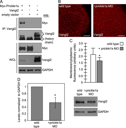
Prickle1a binds Vangl2 and regulates its expression. (A) Western blot (WB) analysis after Vangl2 antibody immunoprecipitation of transfected CHO cell protein extracts. Incubation of protein extracts with Vangl2 antibody co-immunoprecipitates both Vangl2 and Myc-Prickle1a. Whole cell lysates (WCL) demonstrate successful transfection and expression of specific proteins. (B) Confocal images of Vangl2 expression in tailbud stage wild type and prickle1a morphant embryos. Scale bars=20 μm. (C) Quantification of membrane versus cytoplasmic Vangl2 expression; 465 wild type control measurements from three embryos; 742 prickle1a morphant measurements from four embryos (*p<0.0001). (D) Western blot analysis of Vangl2 protein expression normalized to GAPDH in wild type and prickle1a morphant embryos. Bar graph represents data from three independent experiments and the blot depicts results from one of those experiments (*p<0.007). |
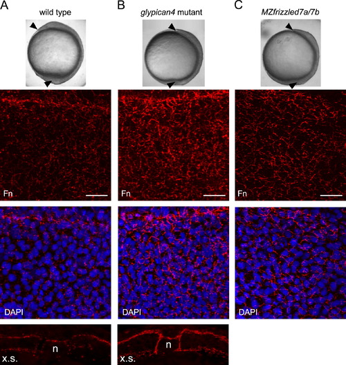
glypican4 and frizzled7 mutant embryos have increased fibronectin assembly. (A–C) Top panels show morphological convergence and extension phenotypes at tailbud stage (see arrowheads) of wild type, glypican4 mutant, and MZfrizzled7a/7b mutant embryos. Middle two panels show confocal images of fibronectin (Fn) immunolabeling without and with nuclear DAPI staining. Scale bars=20 μm. The bottom images in (A) and (B) show Fn expression in cross-sections (x.s.) of tailbud stage embryos (n, notochord). PHENOTYPE:
|
glypican4 and frizzled7 mutant embryos have increased fibronectin assembly. (A–C) Top panels show morphological convergence and extension phenotypes at tailbud stage (see arrowheads) of wild type, glypican4 mutant, and MZfrizzled7a/7b mutant embryos. Middle two panels show confocal images of fibronectin (Fn) immunolabeling without and with nuclear DAPI staining. Scale bars=20 µm. The bottom images in (A) and (B) show Fn expression in cross-sections (x.s.) of tailbud stage embryos (n, notochord). PHENOTYPE:
|
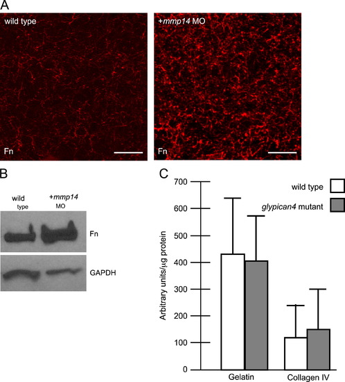
glypican4 mutant embryos have normal proteolytic activity. (A) Confocal images of fibronectin (Fn) expression in tailbud stage wild type and mmp14 morphant embryos. Scale bars=20 μm. (B) Fn and GAPDH western blot of 10 μg total protein extract obtained from wild type and mmp14 morphant embryos. (C) Quantification of proteolytic activity in wild type and glypican4 mutant protein extracts incubated with either gelatin or collagen IV fluorogenic substrates. Here, triplicate experiments were performed using three pools (50 embryos per pool) of total protein prepared from tailbud stage wild type or glypican4 mutant embryos. |
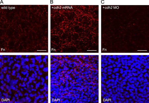
Manipulating Cdh2 expression alters fibronectin fibrillogenesis during gastrulation. (A–C) Confocal images of fibronectin (Fn) expression (without or with nuclear DAPI staining) in wild type, synthetic cdh2 mRNA (200 pg) injected, and cdh2 morphant embryos. Scale bars=20 μm. |
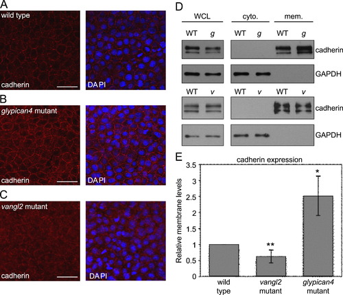
Glypican4 mutant embryos have increased levels of membrane cadherin protein. (A–C) Confocal images of cadherin expression (without or with nuclear DAPI staining) in tailbud stage wild type, glypican4 mutant, and vangl2 mutant embryos. Scale bars=20 μm. (D) Western blot analysis of cadherin expression in embryonic cytoplasmic (cyto.) and membrane (mem.) protein fractions. Cadherin and GAPDH expression in whole cell lysates (WCL) prior to fractionation are also shown. WT, g, and v denote protein extracts obtained from wild type, glypican4 mutant, and vangl2 mutant embryos, respectively. (E) Quantification of western blot fractionation data from triplicate experiments displayed as relative membrane cadherin expression normalized to GAPDH levels in WCL (**p<0.02, *p<0.009). |
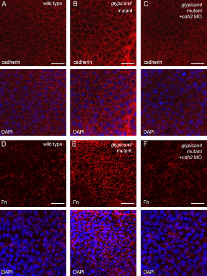
Knockdown of cdh2 suppresses the fibronectin phenotype in glypican4 mutant embryos. Confocal images of cadherin (A–C) or fibronectin (Fn) (D–F) expression (with or without nuclear DAPI staining) in tailbud stage wild type, glypican4 mutant embryos, and glypican4 mutants injected with cdh2 MO. Scale bars=20 &mum. |
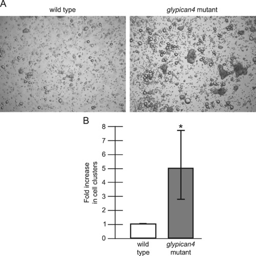
glypican4 mutant embryos have increased intercellular adhesion. (A) Representative 10× images of hanging drop cellular clusters from wild type and glypican4 mutant embryos after repeated pipetting and dissociation. (B) Quantification of the fold increase in cell clusters in glypican4 mutants (*p<0.05). This assay was performed in triplicate, and five 10× images were analyzed per experiment. |
|
|
Reprinted from Developmental Biology, 383(1), Dohn, M.R., Mundell, N.A., Sawyer, L.M., Dunlap, J.A., and Jessen, J.R., Planar cell polarity proteins differentially regulate extracellular matrix organization and assembly during zebrafish gastrulation, 39-51, Copyright (2013) with permission from Elsevier. Full text @ Dev. Biol.