
- Title
-
Functional cooperation of spns2 and fibronectin in cardiac and lower jaw development
- Authors
- Hisano, Y., Ota, S., Takada, S., and Kawahara, A.
- Source
- Full text @ Biol. Open
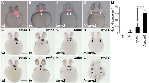
Cardiac morphology.Heart positions are indicated by the arrowheads. (A–D) Cardiac morphology visualized by mRFP expression derived from Tg(cmlc2:mRFP). All images show ventral views at 28hpf. (E–L) Whole-mount in situ hybridization with amhc and vmhc RNA probes. All images show ventral views at 30hpf except for panel L (dorsal view). Genotyping was performed by genomic sequencing after taking pictures, wt (A,E,I), fn mutant (B,F,J), spns2 mutant (C,G,K) and fn;spns2 double mutant (D,H,L). Scale bars: 200μm. (M) Average distances between two hearts from multiple experiments; error bars represent standard deviations. |
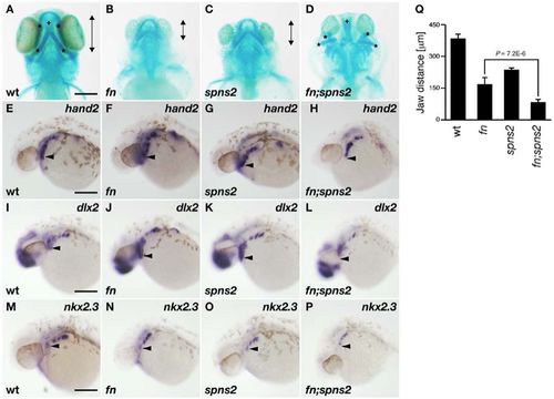
Lower jaw morphology.(A–D) Lower jaw morphology at 4dpf was visualized by Alcian Blue staining (ventral view). Anterior–posterior distances of the ventral pharyngeal arch (*) is indicated by the length of the double-headed arrows. The dorsal pharyngeal structure is identified by the crosses (+). (E–P) Whole-mount in situ hybridization using hand2, dlx2 and nkx2.3 RNA probes. The anteroventral position of these markers is marked by the arrowheads. All images show lateral views at 30hpf. Genotyping was performed by genomic sequencing after taking pictures. wt (A,E,I,M), fn mutant (B,F,J,N), spns2 mutant (C,G,K,O), and fn;spns2 double mutant (D,H,L,P). Scale bars: 200μm. (Q) Average anterior–posterior distances of the ventral pharyngeal arch from multiple experiments; error bars represent standard deviations. |
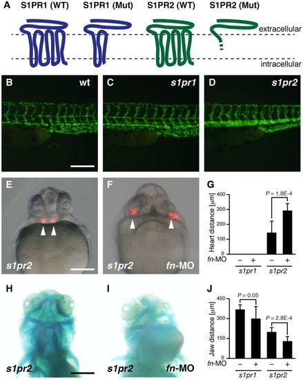
Knockdown phenotype of fibronectin in S1PRs mutant.(A) Membrane topology of S1P receptors and their mutants. The region of frameshift-mediated amino acids compared with the S1PR2 wild type (WT) is shown by the dashed line. (B,C,D) Fluorescence microscopy of intersegmental vessels of wt (B), s1pr1 mutant (C) and s1pr2 mutnat (D) at 2dpf. Endothelial cells are visualized by EGFP expression derived from Tg(fli1a:EGFP). (E,F) Cardiac morphology visualized by mRFP expression derived from Tg(cmlc2:mRFP). All images show ventral views at 28hpf. (G) Average distances between two hearts from multiple experiments; error bars represent standard deviations. (H,I) Lower jaw morphology at 4dpf was visualized by Alcian Blue staining (ventral view). (J) Average anterior–posterior distances of the ventral pharyngeal arch from multiple experiments; error bars represent standard deviations. Genotyping was performed by genomic sequencing or heteroduplex mobility assays after taking pictures. Scale bars: 200μm. |
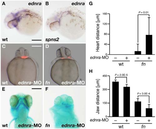
Knockdown phenotype of endothelin receptor A (ednra) in fn mutants.(A,B) Whole-mount in situ hybridization using the ednra RNA probe. The expression of ednra was suppressed in the spns2 mutant. Both images show lateral views at 30hpf. (C,D) Cardiac morphology visualized by mRFP expression derived from Tg(cmlc2:mRFP). Both images show ventral views at 28hpf. (E,F) Lower jaw morphology at 4dpf was visualized by Alcian Blue staining (ventral view). Genotyping was performed by genomic sequencing after taking pictures. wt (A,C,E), spns2 mutant (B) and fn mutant (D,F). Scale bars: 200μm. (G,H) Average distances between hearts (G) and anterior–posterior distances of the ventral pharyngeal arch (H) from multiple experiments; error bars represent standard deviations. |
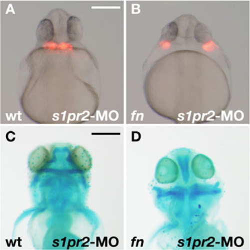
Knockdown phenotype of S1PR2 in fn mutant. (A,B) Cardiac morphology visualized by mRFP expression derived from Tg(cmlc2:mRFP). Both images show ventral views at 28 hpf. (C,D) Lower jaw morphology at 4 dpf was visualized by Alcian Blue staining (ventral view). Genotyping was performed by genomic sequencing after taking pictures. wt (A,C) and fn mutant (B,D). Scale bars: 200 μm. |