
- Title
-
Circulating Bmp10 acts through endothelial Alk1 to mediate flow-dependent arterial quiescence
- Authors
- Laux, D.W., Young, S., Donovan, J.P., Mansfield, C.J., Upton, P.D., and Roman, B.L.
- Source
- Full text @ Development
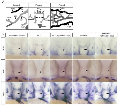
Restoration of alk1 expression does not rescue cxcr4a or edn1 expression in the absence of blood flow. (A) Lateral (anterior left), frontal (anterior upwards) and dorsal (anterior left) views of the zebrafish cranial vasculature at 36 hpf. alk1-positive arteries are light gray, alk1-negative arteries are dark gray and veins are black. alk1-positive arteries are closest to the heart. AA1, aortic arch 1; BA, basilar artery; BCA, basal communicating artery; CaDI, caudal division of the internal carotid artery; ICA, internal carotid artery; LDA, lateral dorsal aorta; PCS, posterior communicating segments; PHBC, primordial hindbrain channel; PMBC, primordial midbrain channel. Scale bar: 50 μm. (B) Whole-mount in situ hybridization for cxcr4a, edn1 and cadherin 5 (cdh5, pan-endothelial control) at 36 hpf. cxcr4a and edn1 are upregulated and downregulated, respectively, in alk1 mutants (column 2) and tnnt2a morphants (column 4) compared with controls (column 1), and expression can be normalized to wild-type/control morphant levels by transgene-mediated, flow-independent alk1 expression in alk1 mutants (column 3) but not in tnnt2a morphants (column 5). cdh5 expression is not altered by alk1 or flow conditions. ne50 for all groups. Frontal views, dorsal upwards. Arrows indicate left CaDI. Scale bar: 50 μm. EXPRESSION / LABELING:
|
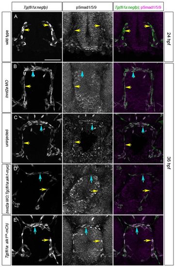
Alk1 activity is dependent on blood flow. (A-E) pSmad1/5/9 (middle column) in endothelial cell nuclei (marked by fli1a:negfp transgene, left column); in merge (right column), EGFP-expressing endothelial cell nuclei are green and pSmad1/5/9 immunofluorescence is magenta. (A) 24 hpf wild type, prior to blood flow; (B) 36 hpf tnnt2a morphant (no flow); (C) 36 hpf wild type; (D) 36 hpf tnnt2a morphant harboring a fli1a:alk1-myc transgene; (E) 36 hpf tnnt2a morphant harboring a fli1a:alk1CA-mCh transgene. Tg(fli1a:alk1CA-mCh) embryos do not have lumenized vessels. In merge (right column), EGFP-expressing endothelial cell nuclei are green, pSmad1/5/9 immunofluorescence is magenta. Yellow and blue arrows indicate endothelial cells in the caudal division of the internal carotid artery (CaDI) and basal communicating artery (BCA), respectively. 2D confocal projections of 50 μm frontal sections, dorsal upwards. Scale bar: 50 μm. See supplementary material Table S1 for fluorescence quantitation. |
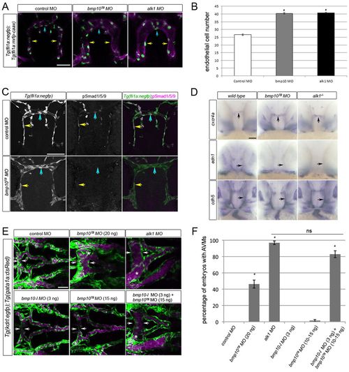
Knockdown of bmp10 phenocopies zebrafish alk1 mutants. (A) CaDIs (caudal divisions of the internal carotid artery; yellow arrows) and BCA (basal communicating artery; blue arrows) in 36 hpf Tg(fli1a:mrfp-caax);Tg(fli1a:negfp) embryos injected with 20 ng control, 20 ng bmp10TB or 2.5 ng alk1 morpholino. Endothelial cell membranes are magenta; nuclei are green. 2D projections of 10 optical sections (Z-step, 2 µm), frontal views, dorsal upwards. Scale bar: 50 μm. (B) Endothelial cell number in the CaDI/BCA in control, bmp10TB and alk1 morphants. n=7-10 in three independent experiments. Values are mean±s.e.m. Student’s t-test: *P<0.001. (C) pSmad1/5/9 (middle column) in endothelial cells (nuclei marked by fli1a:negfp transgene, left column) in 36 hpf control and bmp10TB morphants. In merge (right column), EGFP-expressing endothelial cell nuclei are green, pSmad1/5/9 immunofluorescence is magenta. Yellow and blue arrows indicate endothelial cells in the CaDI and BCA, respectively. 2D confocal projections of 50 μm frontal sections, dorsal upwards. Scale bar: 50 μm. See supplementary material Table S1 for fluorescence quantitation. (D) Whole-mount in situ hybridization for cxcr4a, edn1 and cdh5 (pan-endothelial control) in wild type/control morphant, bmp10TB morphant and alk1-/- at 36 hpf. Frontal views, dorsal upwards. Arrows indicate BCA (vertical) or CaDI (horizontal). Scale bar: 50 μm. (E) Cranial vasculature in 48 hpf Tg(kdrl:gfp);Tg(gata1a:dsRed) embryos injected with bmp10TB morpholino (15-20 ng, as indicated), alk1 morpholino (2.5 ng) and/or bmp10-like morpholino (3 ng). Arrows highlight width of BCA; asterisk indicates AVM. Endothelial cells are green, red blood cells magenta. 2D confocal projections, dorsal views, anterior leftwards. Scale bar: 50 μm. (F) Quantification of AVM development in 48 hpf morpholino-injected embryos. n=43-146. Values are mean±s.e.m. Student’s t-test: *P<0.001; ns, not significant. |
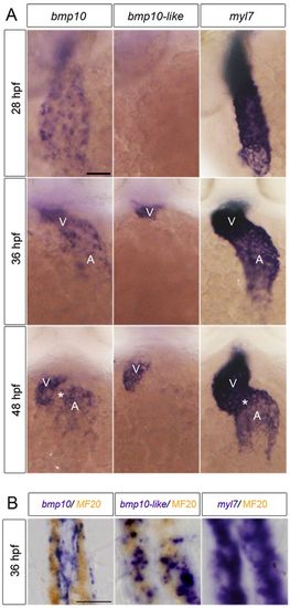
bmp10 and bmp10-like are expressed in the heart. (A) Whole-mount in situ hybridization for bmp10, bmp10-like and myl7 at 28, 36 and 48 hpf. Ventral views, anterior upwards. Scale bar: 50 μm. V, ventricle; A, atrium. Asterisk indicates atrioventricular canal. (B) Sagittal sections (8 μm) through the heart of 36 hpf embryos co-stained with bmp10, bmp10-like or myl7 (purple) and MF20 (sarcomeric myosin; brown). Scale bar: 10 μm. |

Bmp10/Alk1 lies downstream of blood flow in regulation of pSmad1/5/9, cxcr4a and edn1. (A) BRE:luciferase activity in C2C12 cells transfected with human (h) ALK1, zebrafish (z) alk1, hALK1 kinase dead mutant (R411Q), hALK3 (which does not bind BMP10) or lipofectamine (lipo), and treated with rhBMP10. Data are normalized to pTK-Renilla luciferase activity. Values are mean±s.e.m., n=3 biological replicates, each representing 3 technical replicates. (B) pSmad1/5/9 (middle column) in endothelial cells (nuclei marked by fli1a:negfp transgene, left column) of the CaDI (caudal division of the internal carotid artery) of 36 hpf tnnt2a morphants in which endothelial cell alk1 expression [Tg(fli1a:alk1-myc)], BMP10 availability (rhBMP10; 2 nl of 10 μM rhBMP10 injected into base of CaDI at 28 hpf), or both, are restored. In merge (right column), EGFP-expressing endothelial cell nuclei are green, pSmad1/5/9 immunofluorescence is magenta. Yellow arrows indicate endothelial cells in the injected CaDI. 2D confocal projections of 50 μm frontal sections, dorsal upwards. Scale bar: 10 μm. See supplementary material Table S1 for fluorescence quantitation. (C) Whole-mount in situ hybridization for cxcr4a, edn1 and cdh5 (pan-endothelial control) in 36 hpf tnnt2a morphant;Tg(fli1a:alk1-myc) embryos injected at 28 hpf into the base of the left CaDI with 2 nl tracer with or without 10 μM rhBMP10. Yellow arrow indicates injected CaDI, red arrow indicates uninjected CaDI. Asterisk indicates injection site. Frontal views, dorsal upwards; lateral views, right (uninjected) side or left (injected) side. Scale bar: 50 μm. (D) Quantitation of CaDI endothelial cell number in 36 hpf Tg(fli1a:alk1-myc) embryos left uninjected (white bars) or injected at the one-cell stage with tnnt2a morpholino (gray, black bars). At 28 hpf, tnnt2a morphants were injected in the right CaDI with 2 nl tracer (gray bars) or 2 nl 10μM rhBMP10 (black bars). tnnt2a morphants injected intravascularly with tracer exhibited increased endothelial cell number throughout the CaDI/BCA, whereas intravascular injection of rhBMP10 normalized endothelial cell number in the injected CaDI and BCA (basal communicating artery). n=10-12 per group in two independent experiments. Values are mean±s.e.m. Student’s t-test: *P<0.05; **P<0.001; ns, not significant. |
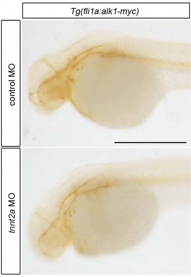
Tg(fli1a:alk1-myc) embryos express Alk1-myc in all endothelial cells in the presence or absence of flow. Immunohistochemistry for myc in 36 hpf Tg(fli1a:alk1-myc) control and tnnt2a morphant embryos. Scale bar: 500 μm. |
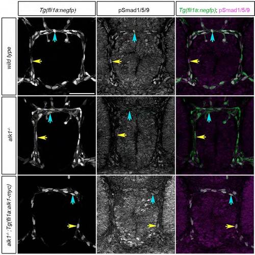
pSmad1/5/9 expression in alk1-positive endothelial cells depends on alk1. pSmad1/5/9 expression (middle column) in endothelial cells (nuclei marked by fli1a:negfp transgene, left column) in wild-type, alk1-/- and alk1-/-;Tg(fli1a:alk1-myc) embryos at 36 hpf. In merge, EGFP-expressing endothelial cell nuclei are green, pSmad1/5/9 immunofluorescence is magenta. Yellow and blue arrows indicate endothelial cells in the CaDI and BCA, respectively. 2D confocal projections of 50 μm frontal sections, dorsal upwards. Scale bar: 50 μm. See Table S1 for fluorescence quantitation. |
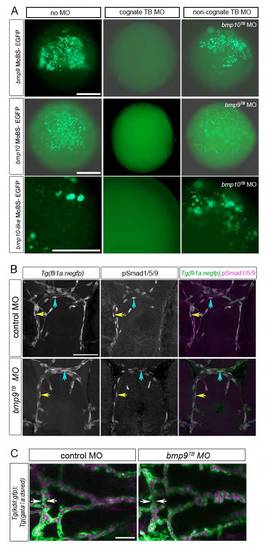
bmp9 knockdown does not phenocopy zebrafish alk1-/- mutants. (A) Translation blocking (TB) morpholino validations. Wild-type embryos injected at the one-cell stage with 50 pg CMV-driven EGFP constructs modified with morpholino-binding sites (MoBS) inserted upstream of the ATG, with or without cognate or non-cognate morpholinos. bmp9TB MO, 7 ng; bmp10TB MO, 20 ng; bmp10-like MO, 3 ng. Embryos were observed at ~6 hpf for EGFP fluorescence. Scale bar: 500 μm (top two rows) or 250 μm (bottom row). (B) pSmad1/5/9 expression (middle column) in endothelial cells (nuclei marked by fli1a:negfp transgene, left column) in 36 hpf embryos injected with 7 ng control or bmp9TB morpholino. Higher amounts of this morpholino were overtly toxic. In merge (right column), EGFP-expressing endothelial cell nuclei are green, pSmad1/5/9 immunofluorescence is magenta. Yellow and blue arrows indicate endothelial cells in the CaDI and BCA, respectively. 2D confocal projections of 50 μm frontal sections, dorsal upwards. Scale bar: 50 μm. See Table S1 for fluorescence quantitation. (C) Cranial vasculature in 48 hpf Tg(kdrl:gfp);Tg(gata1a:dsRed) embryos injected with 7 ng control or bmp9TB morpholino. Arrows highlight width of BCA. Endothelial cells are green, red blood cells are magenta. 2D confocal projections, dorsal views, anterior leftwards. Scale bar: 50 μm. n=207 over five experiments. |
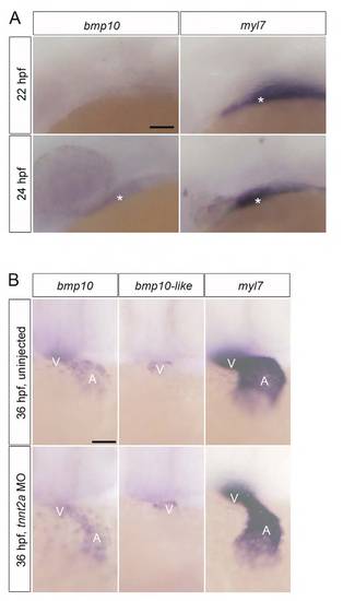
Expression of bmp10 and bmp10-like mRNA does not depend on heartbeat. (A) bmp10 is not expressed at 22 hpf but is faintly expressed in the heart at 24 hpf. Lateral views, anterior leftwards. Scale bar: 100 μm. Asterisk indicates developing heart. (B) bmp10 and bmp10-like expression in control and tnnt2a morphants, which lack heartbeat. Images are representative of n=60 embryos per group. A, atrium; V, ventricle. Ventral views, anterior upwards. Scale bar: 50 μm. myl7 is shown as a pan-myocardial control. EXPRESSION / LABELING:
|
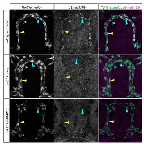
rhBMP10 cannot induce pSmad1/5/9 in the absence of alk1. Intravascular injections of tracer alone or tracer + 2 nl 10 μM rhBMP10 were performed at 28 hpf into wild-type or alk1 mutant embryos. Images show pSmad1/5/9 expression (middle column) in endothelial cells (nuclei marked by fli1a:nEGFP transgene, left column) at 36 hpf. In merge (right column), EGFP-expressing endothelial cell nuclei are green, pSmad1/5/9 immunofluorescence is magenta. Yellow and blue arrows indicate endothelial cells in the CaDI and BCA, respectively. 2D confocal projections of 50 μm frontal sections, dorsal upwards. Scale bar: 50 μm. See Table S1 for fluorescence quantitation. |