
- Title
-
Tissue factor pathway inhibitor-2: A novel gene involved in zebrafish central nervous system development
- Authors
- Zhang, Y., Wang, L., Zhou, W., Wang, H., Zhang, J., Deng, S., Li, W., Li, H., Mao, Z., and Ma, D.
- Source
- Full text @ Dev. Biol.
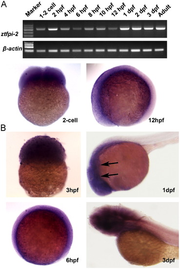
Expression of ztfpi-2 during zebrafish embryogenesis. (A) RT-PCR detection of maternal and zygotic ztfpi-2 transcript from 1-cell stage to adult zebrafish. (B) Pattern of expression of ztfpi-2 as shown using WISH. Stages of development are indicated in the bottom right corner. The expression of ztfpi-2 is indicated by arrows. |
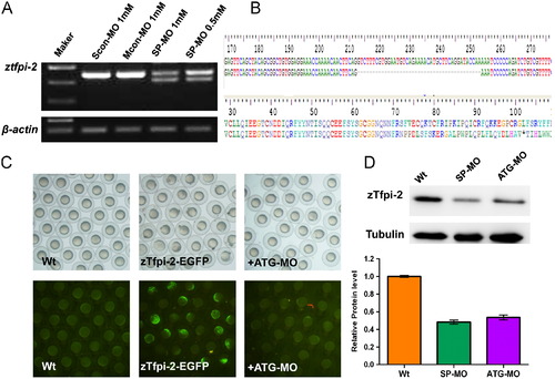
Efficiency of ztfpi-2 knockdown by MOs. (A) The SP-MO altered splicing in zebrafish. The results showed a MO concentration-dependent decrease of ztfpi-2 RNA levels, which indicated that the SP-MO successfully downregulated the expression of ztfpi-2 at 24 hpf. (B) Sequence comparison of ztfpi-2 revealed that 41 bp of exon 2 was lost due to a frameshift mutation after bp 71, leading to a stop codon at bp 102. (C) ATG-MO knockdown of the zTfpi-2-EGFP fusion protein at 8 hpf. Green fluorescence was detectable in a mosaic pattern throughout embryos injected with the zTfpi-2-EGFP plasmid at 38 pg, while GFP expression was absent in nearly all embryos co-injected with 0.6 mM ATG-MO and wild-type embryos without injection. (D) Western blot analysis of zTfpi-2 knockdown efficiency. Embryos injected with 0.5 mM SP-MO or 0.6 mM ATG-MO were harvested at 24 hpf. Tubulin was used as the loading control. Gray scale analysis showed that zTfpi-2 was reduced to 48% of former levels by SP-MO and to 53% by ATG-MO. |
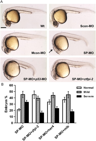
Morphological defects exhibited in ztfpi-2 morphants. (A) SP-MO injected and p53-MO co-injected embryos displayed malformation of the MHB with pigment deficiencies 25 hpf but embryos exposed to Mcon-MO and Scon-MO did not (arrow). These morphological phenotypes were found to be rescued by concomitant injection of ztfpi-2 mRNA. (B) ztfpi-2-deficient embryos were partially rescued by co-injection of 75 pg ztfpi-2, 155 pg her4, and 85 pg mib mRNA. A total of 100 embryos were used per group. Representative results of three independent experiments are shown. PHENOTYPE:
|
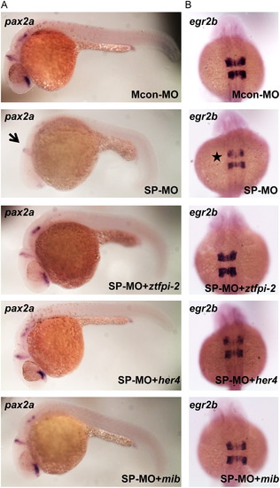
ztfpi-2 regulated the development of the midbrain and hindbrain. All views are lateral. Expression of pax2a in the MHB (A) egr2b in rhombomere 5 (B) at 25 hpf. Embryos injected with 0.5 mM SP-MO displayed weaker expression of pax2a (arrow) and egr2b (asterisk) than those exposed to 0.5 mM Mcon-MO, while wild-type ztfpi-2 RNA was able to rescue their expression in the same region. Also, her4 and mib were able to partially rescue the ztfpi-2 defects in the midbrain and hindbrain. Scale bar: 50 μm. EXPRESSION / LABELING:
|
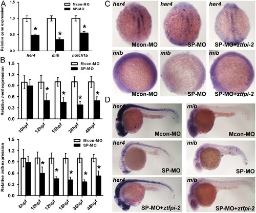
Three members of the Notch pathway were downregulated. (A) The graph shows the results of qPCR analyses of her4, mib and Notch1α mRNA levels when ztfpi-2 was knocked down at 24 hpf. Asterisks indicate a significant difference from the Mcon-MO group (Student′s t test, P<0.05). (B) Results of qPCR showed that both her4 and mib were reduced as early as 3 somites stage. ((C), (D)) WISH indicated that her4 and mib were downregulated while ztfpi-2 mRNA could partially recover their expression at 3 somites stage and 25 hpf. |
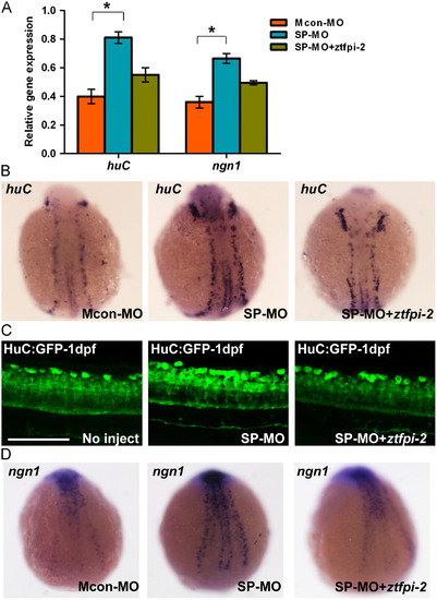
ztfpi-2 morphants exhibited upregulation of huC and ngn1. (A) qPCR analysis showed huC and ngn1 levels have been increased in embryos injected with SP-MO at 25 hpf. *P<0.05. (B) WISH showed, at the 3-somite stage, injected embryos had a higher density of neurons in the neural plate than controls. (C) There were more HuC-positive cells in the SP-MO injected HuC-GFP embryos. A z-stack of fluorescence images was acquired by confocal microscopy at 25 hpf. Scale bar: 100 μm. (D) Dorsal views of Mcon-MO and SP-MO injected embryos showing ngn1 expression at 3 somites stage. The changes could be restored by co-injection of SP-MO with ztfpi-2 mRNA. EXPRESSION / LABELING:
|
ztfpi-2 morphants exhibited upregulation of huC and ngn1. (A) qPCR analysis showed huC and ngn1 levels have been increased in embryos injected with SP-MO at 25 hpf. *P<0.05. (B) WISH showed, at the 3-somite stage, injected embryos had a higher density of neurons in the neural plate than controls. (C) There were more HuC-positive cells in the SP-MO injected HuC-GFP embryos. A z-stack of fluorescence images was acquired by confocal microscopy at 25 hpf. Scale bar: 100 μm. (D) Dorsal views of Mcon-MO and SP-MO injected embryos showing ngn1 expression at 3 somites stage. The changes could be restored by co-injection of SP-MO with ztfpi-2 mRNA. EXPRESSION / LABELING:
PHENOTYPE:
|
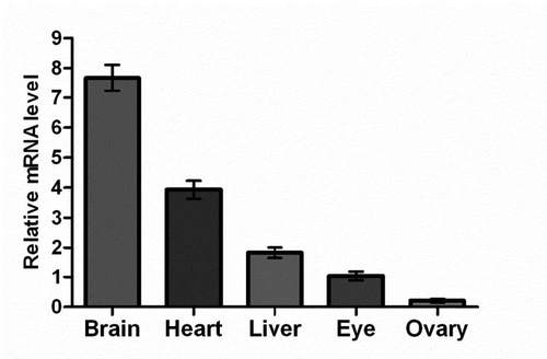
Sites of ztfpi-2 expression in adult zebrafish organs. Expression was most pronounced in the brain and showed a sequential decrease in the heart, liver, eye, and ovary. Values are the mean±SE of four reactions. The mean value of its expression in the eye was arbitrarily set as 1. |
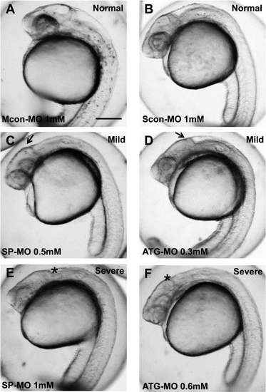
Morphological defects exhibited in ztfpi-2 morphants. Live embryo injection of SP-MO and ATG-MO led to phenotypic defects in the MHB with pigment deficiencies compared with normal at 25 hpf. ((A), (B)) Wild-type without abnormality. ((C), (D)) Mild phenotype between normal and severe (arrow). ((E), (F)) Severe phenotype including lack of the MHB and serious pigment deficiencies (asterisk). Scale bar: 2.0 mm. PHENOTYPE:
|
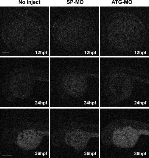
Apoptosis analysis with TUNEL staining of ztfpi-2 morphants at 12 hpf, 24 hpf and 36 hpf. The fluorescent signal for SP-MO and ATG-MO was similar to the uninjected control at all three stages. The scale bar is 200 μm. |
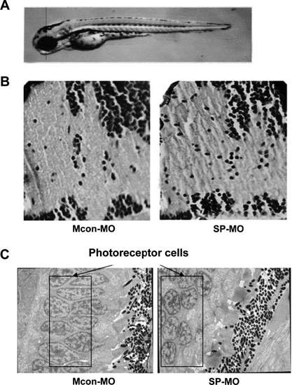
Abnormities of ztfpi-2 knockdown morphants in nerve fibers and retina at 96 hpf. (A) Diagram showing the plane of section. (B) H&E-stained slides of the wild-type and mutant encephalon magnified 100×. Interspaces among the nerve fibers in ztfpi-2 morphants became wider than those in the control. (C) The arrangement of retina photoreceptor cells became disorganized when ztfpi-2 was downregulated by 0.5 mM SPMO (black rectangle). PHENOTYPE:
|
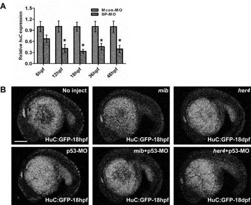
Comparison of the HuC expression between control and morphants. (A) zTfpi-2 knockdown increased the number of neurons in embryos 12 hpf and older stages using qPCR. (B) Ectopic expression of her4 and mib affected HuC expression at 18 hpf. The scale bar is 200 μm. |
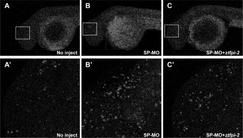
Knockdown of zTfpi-2 increased the neuronal cells. Confocal images of whole-mount immunostaining of HuC/HuD indicated that uninjected embryos (A) had normal HuC/HuD-positive cells, whereas the Tfpi-2 morphants showed increased number of the neuronal cells at 25 hpf. Embryos co-injected with SP-MO and mRNA of ztfpi-2 displayed less severe defects (C). (A2, B2 and C2) are higher magnification of the boxed regions of ((A), (B) and (C)), respectively. |

Unillustrated author statements PHENOTYPE:
|
Reprinted from Developmental Biology, 381(1), Zhang, Y., Wang, L., Zhou, W., Wang, H., Zhang, J., Deng, S., Li, W., Li, H., Mao, Z., and Ma, D., Tissue factor pathway inhibitor-2: A novel gene involved in zebrafish central nervous system development, 38-49, Copyright (2013) with permission from Elsevier. Full text @ Dev. Biol.