
- Title
-
Habenular commissure formation in zebrafish is regulated by the pineal gland-specific gene unc119c
- Authors
- Toyama, R., Kim, M.H., Rebbert, M.L., Gonzales, J., Burgess, H., and Dawid, I.B.
- Source
- Full text @ Dev. Dyn.

Unc119c is involved in HC formation in zebrafish embryos. A, B: Visualizing the HC. Transgenic zebrafish, Tg(AANAT2:eGFP), which express eGFP in the pineal gland, were used. The embryos were fixed at 54 hpf and stained with anti-GFP (green, pineal gland) and anti-acetylated tubulin (red, axonal tracts). The HC is located directly beneath the pineal gland. Dorsal view (A), lateral view (B). C–G: Injection of unc119c MO disrupts HC formation. Wild type zebrafish embryos were injected with 1.5 ng unc119c ATG MO plus 100 pg GFP RNA (C, F), control MO (D), or unc119c ATG MO together with 100 pg unc119c RNA (E, F). Two different unc119c splice MOs, E3I3 and I3E4, also affected HC formation in a dose-dependent manner (G). Statistical significance is indicated as *P < 0.05, and **P < 0.01. Experiments are compared to HC in control embryos (all of which were normal), except for the rescue data (F, second bar, MO+RNA), which are compared to MO-injected embryos (F, first bar). Total number of embryos examined is shown in parentheses above the bars. H: The splice MOs greatly reduced formation of mature mRNA. dvdt, dorsoventral diencephalic tract; hc, habenular commissure; p, pineal gland; pc, posterior commissure. |

Injection of arl3l1 and arl3l2 MO disrupts HC formation. Tg(AANAT2:eGFP) embryos were injected with control (A–C), arl3l1 (D–F, J), or arl3l2 (G–I, K) ATG MO, fixed at 54 hpf and stained with anti-GFP (green, pineal gland) (A, D, and G) and anti-acetylated tubulin (red, neurons) (B, E, and H). C, F, and I are merged images. Arrows indicate the HC (B, E, and H). Dose-dependence of the ATG MO effect (J). K: Splice MOs were injected into wild type embryos, which were stained for acetylated tubulin and classified as above. As the arl3l2 E2I2 MO was somewhat toxic, it was co-injected with 5 ng of p53 MO to suppress cell death (Robu et al., 2007). Experiments were compared to control HC, and statistical significance and number of embryos are indicated as specified in the legend to Figure 1. |
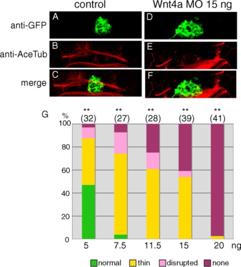
Injection of wnt4 MO affects HC formation in zebrafish embryos. Tg(foxd3:GFP) embryos were used to inject control (A–C) and wnt4a (D–F) MO, fixed at 54 hpf and stained with anti-GFP (green) (A and D) and anti-acetylated tubulin (red) (B and E). C and F are merged images. D–F are rotated counterclockwise by about 20°. G: Dose-dependency of the MO effect. Experiments were compared to control HC, and statistical significance and number of embryos are indicated as specified in the legend to Figure 1. |
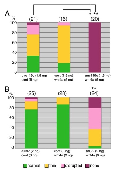
wnt4a MO together with unc119c MO (A) or arl3l2 MO (B) have synergistic effects on HC formation. Tg(AANAT2:eGFP) embryos were injected with MOs at the levels indicated (in ng); cont, control MO. Statistical significance and number of embryos are indicated as specified in the legend to Figure . HC in embryos injected with combined MOs (third bar) were compared to those in single MOinjected embryos (first and second bars). In A each comparison led to different significance, as indicated, while in B both comparisons were highly significant. PHENOTYPE:
|
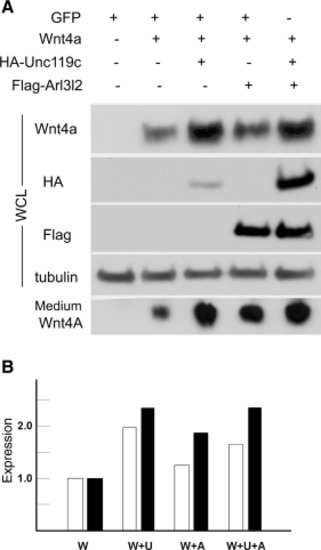
Cultured HEK293T cells were transiently transfected with Wnt4a, with or without HA-Unc119c and Flag-Arl3l2, as described in the Experimental Procedures section. A: Wnt4a, Unc119c, and Arl3l2 in whole cell lysates were detected by immunoblotting, while secreted Wnt4a was recovered from the medium by immunoprecipitation before blotting. B: Quantification of the gel using ImageJ shows the level of Wnt4a extracted from cells (open bars) or recovered from the medium (filled bars). Cells were transfected with Wnt4a (W), Wnt4a and Unc119c (W+U), Wnt4a and Arl3l2 (W+A), or all three (W+U+A). The level of expression in the Wnt4a lane was set as one. |
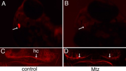
NTR-mediated pineal gland ablation caused HC disruption. Tg(tph2:NfsB-mCherry)y227 embryos were treated with DMSO (control, A, C), or 10 mM Mtz (B, D) for 70 hr, fixed at 78 hpf, and stained with anti-acetylated tubulin (red). Arrows indicate the pineal gland (A and B), or the HC (C and D). EXPRESSION / LABELING:
PHENOTYPE:
|
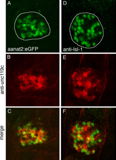
Unc119c antibody visualized Unc119c protein enriched in the pineal gland. Tg(AANAT2:eGFP) embryos were double-stained with anti-GFP (green) to stain photoreceptor cells (A), and unc119c antibody (red, B). Wild type embryos were double-stained with anti-Isl-1(green) to stain projection neurons (D), and Unc119c antibody (E). Merged images (C, F). The pineal gland is outlined in A and D. EXPRESSION / LABELING:
|