
- Title
-
Specification of hepatopancreas progenitors in zebrafish by hnf1ba and wnt2bb
- Authors
- Lancman, J.J., Zvenigorodsky, N., Gates, K.P., Zhang, D., Solomon, K., Humphrey, R.K., Kuo, T., Setiawan, L., Verkade, H., Chi, Y.I., Jhala, U.S., Wright, C.V., Stainier, D.Y., and Dong, P.D.
- Source
- Full text @ Development
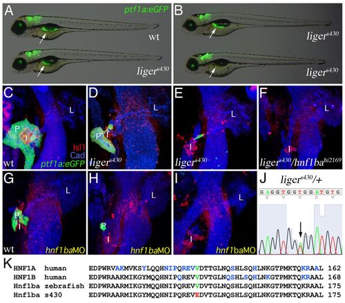
ligers430 is a hypomorphic hnf1ba zebrafish mutant and exhibits MODY5-like pancreas hypoplasia. (A,B) Merged fluorescent/bright-field micrographs of 4 dpf Tg(ptf1a:eGFP)jh1 (arrows indicate pancreas) wild type (A, top) and ligers430 mutants showing the most common severity of pancreas hypoplasia (A, bottom). In ligers430 mutant embryos, the pancreas can be either nearly normal in size (B, top; <10%) or completely absent (B, bottom; <10%). (C-I) Three-dimensional rendering of the foregut endoderm of 80 hpf Tg(ptf1a:eGFP)jh1 wild-type (C), ligers430 (D,E), ligers430/hnf1bahi2169 (F) and hnf1ba-MO injected (G-I) embryos stained for Isl1 and cadherin demonstrating complementation failure by hnf1bahi2169 (F) and phenocopy with Hnf1ba translational knock-down (compare D-F with G-I, respectively). (J) Genomic sequence from a ligers430 heterozygote indicating a molecular lesion (arrow; double peek) in hnf1ba. (K) Amino acid alignment covering part of the atypical POU-specific domain of human HNF1A and HNF1B and wild-type zebrafish and ligers430 Hnf1ba. Red font denotes the valine to glutamic acid substitution at position 147 in ligers430. Blue font indicates amino acids affected by mis-sense mutations in MODY3 and MODY5. PHENOTYPE:
|
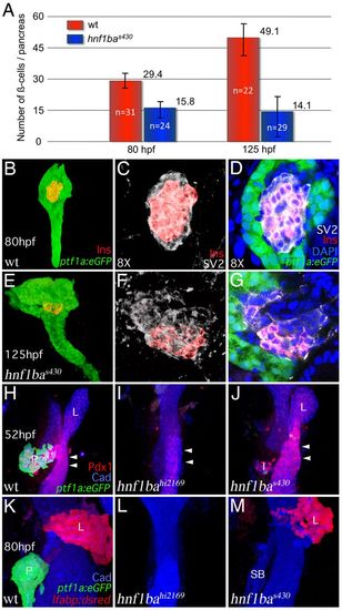
Distinct roles for Hnf1ba in regulating β-cell numbers and ventral pancreas specification. (A) β-Cell nuclei numbers from 80 and 125 hpf wild-type and hnf1bas430 mutants, indicating that at either stage, hnf1bas430 mutants have significantly fewer β-cells. hnf1bas430 mutants (125 hpf) have significantly fewer β-cells than 80 hpf wild-type embryos. Error bars represent s.d. (B-G) Fluorescent confocal microscopy of Tg(ptf1a:eGFP)jh1 80 hpf wild type (B-D) and 125 hpf hnf1bas430 mutant (E-G) pancreas stained for insulin antibodies, SV2 antibodies and DAPI to mark β-cells (red), endocrine cells (white) and nuclei (blue), respectively. Three-dimensional rendering of red and green channels showing an hnf1bas430 mutant at 125 hpf with a larger pancreas (E) than wild-type (B). (C,F) Magnification (8×) of B and E (red and white channels only) to show mildly disorganized islet cells. (D,G) Z-focal plane of C and F (with all channels) demonstrating moderately reduced β-cell number in the hnf1bas430 mutant islet. For a more severe example, see supplementary material Fig. S2. (H-M) Three-dimensional rendering of 52 hpf Tg(ptf1a:eGFP)jh1 (pancreas, green) foregut endoderm in wild-type (H), hnf1bahi2169 mutants (I) and hnf1bas430 embryos (J) stained for Pdx1 to mark the duodenal intestine and cadherin to mark the endoderm epithelium. In contrast to hnf1bahi2169 (I), Pdx1 expression (arrowheads) in the intestine and dorsal pancreatic islet (I) is not lost in hnf1bas430 embryos (J). (K-M) Three-dimensional rendering of 80 hpf Tg(ptf1a:eGFP)jh1; Tg(lfabp:dsRed)gz2 (pancreas, green; liver, red) foregut endoderm in wild-type (K) hnf1bahi2169 (L) and hnf1bas430 embryos (M) stained for cadherin showing specific loss of ventral pancreas in hnf1bas430 hypomorphic mutants. Unlike the hnf1bahi2169 (L), liver (L) and swimbladder (SB) are not lost in the hnf1bas430 embryos. EXPRESSION / LABELING:
|

hnf1ba and wnt2bb cooperate to specify the hepatopancreas organ system after mid-somitogenesis. (A-F) Three-dimensional rendering of 80 hpf Tg(ptf1a:eGFP)jh1; Tg(lfabp:dsRed)gz2 (pancreas, green; liver, red) foregut endoderm in wild type (A), wnt2bbs404 mutant (B), hnf1bas430 mutant (C) and wnt2bbs404; hnf1bas430 double mutants (D-F) stained for cadherin (epithelial endoderm) and with monoclonal antibody 2F11 to mark extra hepatopancreas ducts (*) (dashed outline). Relative to wild type, the wnt2bbs404 mutant liver (L) and the hnf1bas430 mutant pancreas (P) are reduced in size. The wnt2bbs404; hnf1bas430 double mutant liver and pancreas (arrow) are more severely reduced, or lost. Double mutants can exhibit complete loss of the hepatopancreas system (E,F; white arrowheads). (G,H) Three-dimensional rendering of 80 hpf Tg(ptf1a:eGFP)jh1 (pancreas, green) foregut endoderm in wild-type (G) and wnt2bbs404; hnf1bas430 double mutants (H) stained for cadherin and Isl1 to mark the hepatopancreas mesenchyme (brackets) and islet (I). Double mutants lacking the entire hepatopancreas system still develop Isl1+ mesenchyme and islet (yellow arrowheads indicate the swimbladder). (I) Merged fluorescent/brightfield micrographs of 4 dpf Tg(ptf1a:eGFP)jh1; Tg(lfabp:dsRed)gz2 wnt2bbs404 mutant (top) and wnt2bbs404; hnf1bas430 double mutants (bottom) showing no obvious body phenotypes other than liver and pancreas agenesis (arrows). (J,K) Three-dimensional rendering of 80 hpf foregut endoderm, heat-shock-induced Tg(hsp70l:dkk1-GFP)w32; Tg(ptf1a:eGFP)jh1; Tg(lfabp:dsRed)gz2 and Tg(hsp70l:dkk1-GFP)w32; hnf1bas430; Tg(ptf1a:eGFP)jh1; Tg(lfabp:dsRed)gz2 stained for cadherin and with 2F11. Heat-shock-induced expression of dkk1-GFP in wild-type background does not lead to liver or pancreas agenesis (J), whereas in the hnf1bas430 mutant background, the hepatopancreas system is completely lost (K) (white arrow). The swimbladder (yellow arrowhead) is not lost in wnt2bbs404; hnf1bas430 double mutants or in heat-shock-induced Tg(hsp70l:dkk1-GFP)w32; hnf1bas430 mutants (D-F,H,K). PHENOTYPE:
|
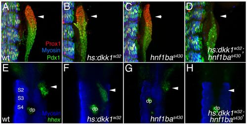
Specification of hepatopancreas progenitors by Hnf1ba and Wnt signaling. (A-D) 3D rendering (lateral view showing somites for positional reference) of 36 hpf foregut endoderm in wild-type (A), Tg(hsp70l:dkk1-GFP)w32 (B), hnf1bas430 (C) and Tg(hsp70l:dkk1-GFP)w32; hnf1bas430 embryos (D) stained for Prox1, Pdx1 and myosin (somites). Relative to wild type, Prox1 foregut expression (arrowheads) is similar or mildly reduced in both Tg(hsp70l:dkk1-GFP)w32 and hnf1bas430 embryos. Prox1 expression is severely reduced or lost in Tg(hsp70l:dkk1-GFP)w32; hnf1bas430 embryos. (E-G) Double in situ hybridization and antibody staining (ventral view) for the early hepatopancreas progenitor marker hhex and myosin at 36 hpf. hhex is expressed (arrowheads) in the foregut endoderm and dorsal pancreas (dp) in wild type (E) but reduced in both the Tg(hsp70l:dkk1-GFP)w32 (F) and the hnf1bas430 (G) embryos. (H) hhex expression (arrowhead) is undetectable in the foregut endoderm of Tg(hsp70l:dkk1-GFP)w32; hnf1bas430 embryos. |
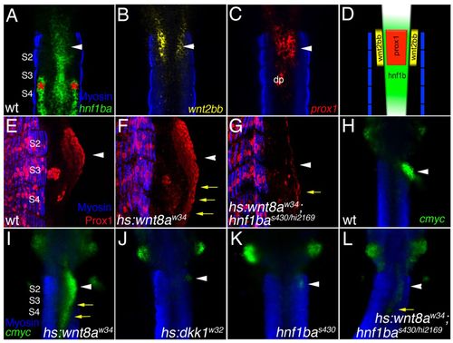
Hnf1ba is required for Wnt signaling activity in the foregut endoderm. (A-C) Double antibody (myosin; blue) and in situ hybridization staining (ventral view) of 24 hpf wild-type foregut endoderm for expression of hnf1ba (A), wnt2bb (B) and prox1 (C, arrowheads). (D) Illustration summarizing foregut expression domains showing restricted prox1 expression within the anterior region of the hnf1ba foregut expression domain and adjacent to the wnt2bb expression domain in the lateral plate mesoderm. Pronephric hnf1ba expression (red asterisks in A) and dorsal pancreas prox1 expression (dp) are not depicted in the illustration. (E-G) Three-dimensional rendering (lateral view) of 36 hpf foregut endoderm in wild-type (E), Tg(hsp70l:wnt8a-GFP)w34 (F) and Tg(hsp70l:wnt8a-GFP)w34; hnf1bas430/hi2169 (G) embryos heat-shocked from 21-28 hpf at 37°C and stained for Prox1 and myosin. Compared with wild type, heat-shocked Tg(hsp70l:wnt8a-GFP)w34 embryos show significant posterior expansion of Prox1 in the foregut endoderm (yellow arrows; n=8/8). In Tg(hsp70l:wnt8a-GFP)w34; hnf1bas430/hi2169 embryos, only weak or no Prox1 expression is observed (yellow arrows; n=6/6). (H-L) Double antibody and in situ hybridization staining (ventral view) of cmyc at 36 hpf in wild-type (H), Tg(hsp70l:wnt8a-GFP)w34 (I), Tg(hsp70l:dkk1-GFP)w32 (J), hnf1bas430-/- (K) and Tg(hsp70l:dkk1-GFP)w32; hnf1bas430/hi216 (L) embryos heat-shocked from 21 to 28 hpf. cmyc expression is restricted in the wild-type foregut endoderm (white arrowhead in H) but is expanded posteriorly in Tg(hsp70l:wnt8a-GFP)w34 (yellow arrows in I; n=12/12) or severely reduced in Tg(hsp70l:dkk1-GFP)w32 (white arrowhead in J) embryos. cmyc expression is reduced or lost in the hnf1bas430 foregut endoderm (white arrowhead in K; n=4/4), and only weakly induced in Tg(hsp70l:wnt8a-GFP)w34; hnf1bas430/hi2169 embryos (yellow arrow in L; n=4/4). |
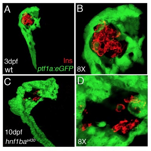
Severe example of β-cell number reduction and disorganization in a hnf1bas430 mutant with mild exocrine hypoplasia. (A-D) Fluorescent confocal microscopy of Tg(ptf1a:eGFP)jh1 (green) 3 dpf wild-type (A,B) and 10 dpf hnf1bs430 mutant (C,D) pancreas stained for insulin to mark β-cells (red). 3D rendering showing an hnf1bas430 mutant at 10 dpf with a larger exocrine pancreas (C) than that of a wild type at 3 dpf (A). (B,D) Magnification (8×) of A and C showing that β-cells (red) remain both reduced and disorganized in hnf1bas430 mutants at 10 dpf. PHENOTYPE:
|

Unillustrated author statements |