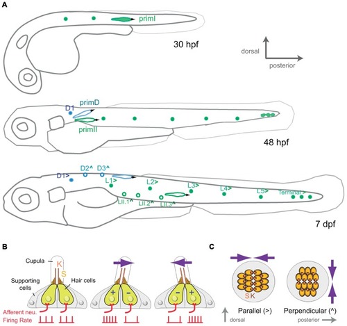
Development and organization of the posterior lateral-line sensory receptors. (A) Lateral view of a developing zebrafish at around 30, 48 hpf and 7 dpf (days post-fertilization) showing the development of the mechanoreceptive neuromasts that form the posterior lateral line. primI, first primordium; primII, second primordium; primD, dorsal primordium. (>) and (∧) indicate parallel and perpendicular neuromasts, respectively. (B) Lateral view of a neuromast. The hair-bundle comprises the kinocilium (K) and the stereocilia (S), and is contained within a gelatinous cupula. A neuromast contains two populations of hair cells of opposing hair-bundle polarities. Thus, a water movement (blue arrow) bending the cupula in a given direction depolarizes (+) one population of hair cells, whereas hyperpolarizes (-) the other one. This is translated into an increase or a decrease of the firing rate of the afferent neuron associated to each hair-cell population. (C) Top view of a parallel and a perpendicular neuromast, which are sensitive to water movements across orthogonal axes.
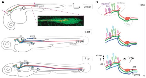
Assembly of the posterior lateral-line neural map. (A) Lateral view of a developing zebrafish at around 30 hpf, 3 and 7 dpf showing the coincident progressive development of the lateralis afferents and their peripheral targets. Arrows at 30 hpf indicate growing lateralis afferent’s central and peripheral axons. PLLg: posterior lateralis ganglion formed by the lateralis afferent’s cell bodies. (A’) A single lateralis afferent neuron is labeled in red in a transgenic zebrafish embryo expressing GFP in primI. The peripheral axonal growth cone can be observed within the migrating primordium. (B) Lateral view of the developing hindbrain and lateralis afferent’s central axons at around 30 hpf, 3 and 7 dpf depicting an hypothetical temporal code that matches lateralis afferents with second-order neurons that are born at similar times. Both in (A) and (B), red and green lateralis afferents belong to the first neuronal subclass which projects dorsal axons that contact the Mauthner cell. Red and green neurons innervate posterior and anterior primary neuromasts, respectively. Blue lateralis afferents belong to the second neuronal subclass which projects ventrolateral axons that do not contact the Mauthner cell. The two neuronal subclasses form a dimorphic neural map (DI) whereas only the neurons of the first subclass shape the somatotopic map (S). Neurons in red are the first-born neurons whereas neurons in blue are the latest-born neurons. primI, first primordium; primD, dorsal primordium; primII, second primordium. L1, Terminal and D1 neuromasts are primary and parallel (>). LII.1 and D2 neuromasts are secondary and perpendicular (∧).
Acknowledgments
This image is the copyrighted work of the attributed author or publisher, and
ZFIN has permission only to display this image to its users.
Additional permissions should be obtained from the applicable author or publisher of the image.
Full text @ Front. Neural Circuits
Your Input Welcome
Thank you for submitting comments. Your input has been emailed to ZFIN curators who may contact you if
additional information is required.
Oops. Something went wrong. Please try again later.